This entry represents the z-alpha domain found in double-stranded RNA-specific adenosine deaminase (ADAR), an RNA-editing enzyme. The z-alpha domain is a Z-DNA binding domain [ (PUBMED:10535945) ]. This domain can also be found in Z-DNA-binding protein 1, a cytosolic DNA sensor that activates the innate immune system [ (PUBMED:17618271) ].
Family alignment:
There are 934 Zalpha domains in 478 proteins in SMART's nrdb database.
Click on the following links for more information.
Evolution (species in which this domain is found)
Taxonomic distribution of proteins containing Zalpha domain.
This tree includes only several representative species. The complete taxonomic breakdown of all proteins with Zalpha domain is also avaliable.
Click on the protein counts, or double click on taxonomic names to display all proteins containing Zalpha domain in the selected taxonomic class.
Literature (relevant references for this domain)
Primary literature is listed below; Automatically-derived, secondary literature is also avaliable.
The solution structure of the Zalpha domain of the human RNA editing enzyme ADAR1 reveals a prepositioned binding surface for Z-DNA.
Proc Natl Acad Sci U S A. 1999; 96: 12465-70
Display abstract
Double-stranded RNA deaminase I (ADAR1) contains the Z-DNA binding domain Zalpha. Here we report the solution structure of free Zalpha and map the interaction surface with Z-DNA, confirming roles previously assigned to residues by mutagenesis. Comparison with the crystal structure of the (Zalpha)(2)/Z-DNA complex shows that most Z-DNA contacting residues in free Zalpha are prepositioned to bind Z-DNA, thus minimizing the entropic cost of binding. Comparison with homologous (alpha+beta)helix-turn-helix/B-DNA complexes suggests that binding of Zalpha to B-DNA is disfavored by steric hindrance, but does not eliminate the possibility that related domains may bind to both B- and Z-DNA.
Crystal structure of the Zalpha domain of the human editing enzyme ADAR1 bound to left-handed Z-DNA.
Science. 1999; 284: 1841-5
Display abstract
The editing enzyme double-stranded RNA adenosine deaminase includes a DNA binding domain, Zalpha, which is specific for left-handed Z-DNA. The 2.1 angstrom crystal structure of Zalpha complexed to DNA reveals that the substrate is in the left-handed Z conformation. The contacts between Zalpha and Z-DNA are made primarily with the "zigzag" sugar-phosphate backbone, which provides a basis for the specificity for the Z conformation. A single base contact is observed to guanine in the syn conformation, characteristic of Z-DNA. Intriguingly, the helix-turn-helix motif, frequently used to recognize B-DNA, is used by Zalpha to contact Z-DNA.
The Zalpha domain from human ADAR1 binds to the Z-DNA conformer of many different sequences.
Nucleic Acids Res. 1998; 26: 3486-93
Display abstract
Z-DNA, the left-handed conformer of DNA, is stabilized by the negative supercoiling generated during the movement of an RNA polymerase through a gene. Recently, we have shown that the editing enzyme ADAR1 (double-stranded RNA adenosine deaminase, type 1) has two Z-DNA binding motifs, Zalpha and Zbeta, the function of which is currently unknown. Here we show that a peptide containing the Zalpha motif binds with high affinity to Z-DNA as a dimer, that the binding site is no larger than 6 bp and that the Zalpha domain can flip a range of sequences, including d(TA)3, into the Z-DNAconformation. Evidence is also presented to show that Zalpha and Zbeta interact to form a functional DNA binding site. Studies with atomic force microscopy reveal that binding of Zalpha to supercoiled plasmids is associated with relaxation of the plasmid. Pronounced kinking of DNA is observed, and appears to be induced by binding of Zalpha. The results reported here support a model where the Z-DNA binding motifs target ADAR1 to regions of negative supercoiling in actively transcribing genes. In this situation, binding by Zalpha would be dependent upon the local level of negative superhelicity rather than the presence of any particular sequence.
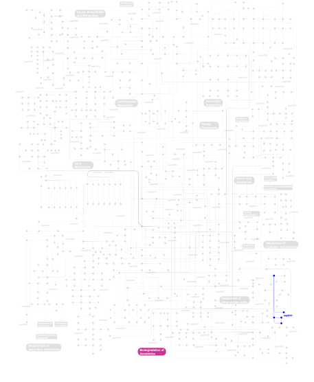
Metabolism (metabolic pathways involving proteins which contain this domain)
Click the image to view the interactive version of the map in iPath
This information is based on mapping of SMART genomic protein database to KEGG orthologous groups. Percentage points are related to the number of proteins with Zalpha domain which could be assigned to a KEGG orthologous group, and not all proteins containing Zalpha domain. Please note that proteins can be included in multiple pathways, ie. the numbers above will not always add up to 100%.