The domain within your query sequence starts at position 1727 and ends at position 1862; the E-value for the LamG domain shown below is 2.33e-23.
KDFEISLKFQTDQLNGLLLFIHNTEGPDFLAVELKRGLLSFKFNSSLVFTRVDLRLGLAD
CDGKWNTVSIKKEGSVVSVRVNALKKSTSQAGGQPLLVNSPVYLGGIPRELQDAYRHLTL
EPGFRGCVKEVAFARG
LamGLaminin G domain |
|---|
| SMART accession number: | SM00282 |
|---|---|
| Description: | - |
| Family alignment: |
There are 74121 LamG domains in 31952 proteins in SMART's nrdb database.
Click on the following links for more information.
- Evolution (species in which this domain is found)
-
Taxonomic distribution of proteins containing LamG domain.
This tree includes only several representative species. The complete taxonomic breakdown of all proteins with LamG domain is also avaliable.
Click on the protein counts, or double click on taxonomic names to display all proteins containing LamG domain in the selected taxonomic class.
- Literature (relevant references for this domain)
-
Primary literature is listed below; Automatically-derived, secondary literature is also avaliable.
- Beckmann G, Hanke J, Bork P, Reich JG
- Merging extracellular domains: fold prediction for laminin G-like and amino-terminal thrombospondin-like modules based on homology to pentraxins.
- J Mol Biol. 1998; 275: 725-30
- Display abstract
Using a new method for construction and database searches of sequence consensus strings, we have identified a new superfamily of protein modules comprising laminin G, thrombospondin N and the pentraxin families. The conserved patterns correspond mainly to hydrophobic core residues located in central beta strands of the known three-dimensional structures of two pentraxins, the human C-reactive protein and the serum amyloid P-component. Thus, we predict a similar jellyroll fold for all members of this superfamily. In addition, the conservation of two exposed aspartate residues in the majority of superfamily members suggests hitherto unrecognised functional sites.
- Disease (disease genes where sequence variants are found in this domain)
-
SwissProt sequences and OMIM curated human diseases associated with missense mutations within the LamG domain.
Protein Disease Laminin subunit alpha-2 (P24043) (SMART) OMIM:156225: Muscular dystrophy, congenital merosin-deficient - Metabolism (metabolic pathways involving proteins which contain this domain)
-

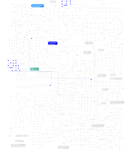
Click the image to view the interactive version of the map in iPath% proteins involved KEGG pathway ID Description 27.86 map04512 ECM-receptor interaction 22.39 map04510 Focal adhesion 14.43 map05222 Small cell lung cancer 12.44 map04514 Cell adhesion molecules (CAMs) 8.46 map04360 Axon guidance 3.98 map05060 Prion disease 2.99 map04610 Complement and coagulation cascades 1.99 map00511 N-Glycan degradation 1.99  map00600
Sphingolipid metabolism 1.99 map01032 Glycan structures - degradation 0.50  map00562
Inositol phosphate metabolism 0.50 map04140 Regulation of autophagy 0.50 map04070 Phosphatidylinositol signaling system This information is based on mapping of SMART genomic protein database to KEGG orthologous groups. Percentage points are related to the number of proteins with LamG domain which could be assigned to a KEGG orthologous group, and not all proteins containing LamG domain. Please note that proteins can be included in multiple pathways, ie. the numbers above will not always add up to 100%.
- Structure (3D structures containing this domain)
3D Structures of LamG domains in PDB
PDB code Main view Title 1c4r 
THE STRUCTURE OF THE LIGAND-BINDING DOMAIN OF NEUREXIN 1BETA: REGULATION OF LNS DOMAIN FUNCTION BY ALTERNATIVE SPLICING 1d2s 
CRYSTAL STRUCTURE OF THE N-TERMINAL LAMININ G-LIKE DOMAIN OF SHBG IN COMPLEX WITH DIHYDROTESTOSTERONE 1dyk 
Laminin alpha 2 chain LG4-5 domain pair 1f5f 
CRYSTAL STRUCTURE OF THE N-TERMINAL G-DOMAIN OF SHBG IN COMPLEX WITH ZINC 1h30 
C-terminal LG domain pair of human Gas6 1kdk 
THE STRUCTURE OF THE N-TERMINAL LG DOMAIN OF SHBG IN CRYSTALS SOAKED WITH EDTA 1kdm 
THE CRYSTAL STRUCTURE OF THE HUMAN SEX HORMONE-BINDING GLOBULIN (TETRAGONAL CRYSTAL FORM) 1lhn 
CRYSTAL STRUCTURE OF THE N-TERMINAL LG-DOMAIN OF SHBG IN COMPLEX WITH 5ALPHA-ANDROSTANE-3BETA,17ALPHA-DIOL 1lho 
CRYSTAL STRUCTURE OF THE N-TERMINAL LG-DOMAIN OF SHBG IN COMPLEX WITH 5ALPHA-ANDROSTANE-3BETA,17BETA-DIOL 1lhu 
CRYSTAL STRUCTURE OF THE N-TERMINAL LG-DOMAIN OF SHBG IN COMPLEX WITH ESTRADIOL 1lhv 
CRYSTAL STRUCTURE OF THE N-TERMINAL LG-DOMAIN OF SHBG IN COMPLEX WITH NORGESTREL 1lhw 
CRYSTAL STRUCTURE OF THE N-TERMINAL LG-DOMAIN OF SHBG IN COMPLEX WITH 2-METHOXYESTRADIOL 1okq 
LAMININ ALPHA 2 CHAIN LG4-5 DOMAIN PAIR, CA1 SITE MUTANT 1pz7 
Modulation of agrin function by alternative splicing and Ca2+ binding 1pz8 
Modulation of agrin function by alternative splicing and Ca2+ binding 1pz9 
Modulation of agrin function by alternative splicing and Ca2+ binding 1q56 
NMR structure of the B0 isoform of the agrin G3 domain in its Ca2+ bound state 1qu0 
CRYSTAL STRUCTURE OF THE FIFTH LAMININ G-LIKE MODULE OF THE MOUSE LAMININ ALPHA2 CHAIN 1sli 
LEECH INTRAMOLECULAR TRANS-SIALIDASE COMPLEXED WITH DANA 1sll 
SIALIDASE L FROM LEECH MACROBDELLA DECORA 2c5d 
Structure of a minimal Gas6-Axl complex 2h0b 
Crystal Structure of the second LNS/LG domain from Neurexin 1 alpha 2jd4 
Mouse laminin alpha1 chain, domains LG4-5 2r16 
Crystal Structure of bovine neurexin 1 alpha LNS/LG domain 4 (with no splice insert) 2r1b 
Crystal Structure of rat neurexin 1beta with a splice insert at SS#4 2r1d 
Crystal structure of rat neurexin 1beta in the Ca2+ containing form 2sli 
LEECH INTRAMOLECULAR TRANS-SIALIDASE COMPLEXED WITH 2,7-ANHYDRO-NEU5AC, THE REACTION PRODUCT 2wjs 
Crystal structure of the LG1-3 region of the laminin alpha2 chain 2wqz 
Crystal structure of synaptic protein neuroligin-4 in complex with neurexin-beta 1: alternative refinement 2xb6 
Revisited crystal structure of Neurexin1beta-Neuroligin4 complex 3asi 
Alpha-Neurexin-1 ectodomain fragment; LNS5-EGF3-LNS6 3b3q 
Crystal structure of a synaptic adhesion complex 3biw 
Crystal structure of the Neuroligin-1/Neurexin-1beta synaptic adhesion complex 3bod 
Structure of mouse beta-neurexin 1 3bop 
Structure of mouse beta-neurexin 2D4 3mw2 
Crystal structure of beta-neurexin 1 with the splice insert 4 3mw3 
Crystal structure of beta-neurexin 2 with the splice insert 4 3mw4 
Crystal structure of beta-neurexin 3 without the splice insert 4 3poy 
Crystal Structure of the alpha-Neurexin-1 ectodomain, LNS 2-6 3pve 
Crystal structure of the G2 domain of Agrin from Mus Musculus 3qcw 
Structure of neurexin 1 alpha (domains LNS1-LNS6), no splice inserts 3r05 
Structure of neurexin 1 alpha (domains LNS1-LNS6), with splice insert SS3 3sh4 
Laminin G like domain 3 from human perlecan 3sh5 
Calcium-bound Laminin G like domain 3 from human perlecan 3sli 
LEECH INTRAMOLECULAR TRANS-SIALIDASE COMPLEXED WITH 2,7-ANHYDRO-NEU5AC PREPARED BY SOAKING WITH 3'-SIALYLLACTOSE 3v64 
Crystal Structure of agrin and LRP4 3v65 
Crystal structure of agrin and LRP4 complex 3vkf 
Crystal Structure of Neurexin 1beta/Neuroligin 1 complex 4c1w 
4C1W 4c1x 
4C1X 4qvs 
4QVS 4ra0 
4RA0 4sli 
LEECH INTRAMOLECULAR TRANS-SIALIDASE COMPLEXED WITH 2-PROPENYL-NEU5AC, AN INACTIVE SUBSTRATE ANALOGUE 4zxk 
4ZXK 5ik4 
5IK4 5ik5 
5IK5 5ik7 
5IK7 5ik8 
5IK8 - Links (links to other resources describing this domain)
-
PFAM laminin_G

