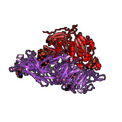
| PDB code | Main view | Title | | 1ao3 |  | A3 DOMAIN OF VON WILLEBRAND FACTOR |
| 1aox |  | I DOMAIN FROM INTEGRIN ALPHA2-BETA1 |
| 1atz |  | HUMAN VON WILLEBRAND FACTOR A3 DOMAIN |
| 1auq |  | A1 DOMAIN OF VON WILLEBRAND FACTOR |
| 1bho |  | MAC-1 I DOMAIN MAGNESIUM COMPLEX |
| 1bhq |  | MAC-1 I DOMAIN CADMIUM COMPLEX |
| 1ck4 |  | CRYSTAL STRUCTURE OF RAT A1B1 INTEGRIN I-DOMAIN. |
| 1cqp |  | CRYSTAL STRUCTURE ANALYSIS OF THE COMPLEX LFA-1 (CD11A) I-DOMAIN / LOVASTATIN AT 2.6 A RESOLUTION |
| 1dgq |  | NMR SOLUTION STRUCTURE OF THE INSERTED DOMAIN OF HUMAN LEUKOCYTE FUNCTION ASSOCIATED ANTIGEN-1 |
| 1dzi |  | integrin alpha2 I domain / collagen complex |
| 1fe8 |  | CRYSTAL STRUCTURE OF THE VON WILLEBRAND FACTOR A3 DOMAIN IN COMPLEX WITH A FAB FRAGMENT OF IGG RU5 THAT INHIBITS COLLAGEN BINDING |
| 1fns |  | CRYSTAL STRUCTURE OF THE VON WILLEBRAND FACTOR (VWF) A1 DOMAIN I546V MUTANT IN COMPLEX WITH THE FUNCTION BLOCKING FAB NMC4 |
| 1idn |  | MAC-1 I DOMAIN METAL FREE |
| 1ido |  | I-DOMAIN FROM INTEGRIN CR3, MG2+ BOUND |
| 1ijb |  | The von Willebrand Factor mutant (I546V) A1 domain |
| 1ijk |  | The von Willebrand Factor mutant (I546V) A1 domain-botrocetin Complex |
| 1jeq |  | Crystal Structure of the Ku Heterodimer |
| 1jey |  | Crystal Structure of the Ku heterodimer bound to DNA |
| 1jlm |  | I-DOMAIN FROM INTEGRIN CR3, MN2+ BOUND |
| 1jv2 |  | CRYSTAL STRUCTURE OF THE EXTRACELLULAR SEGMENT OF INTEGRIN ALPHAVBETA3 |
| 1l5g |  | CRYSTAL STRUCTURE OF THE EXTRACELLULAR SEGMENT OF INTEGRIN AVB3 IN COMPLEX WITH AN ARG-GLY-ASP LIGAND |
| 1lfa |  | CD11A I-DOMAIN WITH BOUND MN++ |
| 1m10 |  | Crystal structure of the complex of Glycoprotein Ib alpha and the von Willebrand Factor A1 Domain |
| 1m1u |  | AN ISOLEUCINE-BASED ALLOSTERIC SWITCH CONTROLS AFFINITY AND SHAPE SHIFTING IN INTEGRIN CD11B A-DOMAIN |
| 1m1x |  | CRYSTAL STRUCTURE OF THE EXTRACELLULAR SEGMENT OF INTEGRIN ALPHA VBETA3 BOUND TO MN2+ |
| 1m2o |  | Crystal Structure of the Sec23-Sar1 complex |
| 1m2v |  | Crystal Structure of the yeast Sec23/24 heterodimer |
| 1mf7 |  | INTEGRIN ALPHA M I DOMAIN |
| 1mhp |  | Crystal structure of a chimeric alpha1 integrin I-domain in complex with the Fab fragment of a humanized neutralizing antibody |
| 1mjn |  | Crystal Structure of the intermediate affinity aL I domain mutant |
| 1mq8 |  | Crystal structure of alphaL I domain in complex with ICAM-1 |
| 1mq9 |  | Crystal structure of high affinity alphaL I domain with ligand mimetic crystal contact |
| 1mqa |  | Crystal structure of high affinity alphaL I domain in the absence of ligand or metal |
| 1n3y |  | Crystal structure of the alpha-X beta2 integrin I domain |
| 1n9z |  | INTEGRIN ALPHA M I DOMAIN MUTANT |
| 1na5 |  | INTEGRIN ALPHA M I DOMAIN |
| 1oak |  | CRYSTAL STRUCTURE OF THE VON WILLEBRAND FACTOR (VWF) A1 DOMAIN IN COMPLEX WITH THE FUNCTION BLOCKING NMC-4 FAB |
| 1pt6 |  | I domain from human integrin alpha1-beta1 |
| 1q0p |  | A domain of Factor B |
| 1qc5 |  | I Domain from Integrin Alpha1-Beta1 |
| 1qcy |  | THE CRYSTAL STRUCTURE OF THE I-DOMAIN OF HUMAN INTEGRIN ALPHA1BETA1 |
| 1rd4 |  | An allosteric inhibitor of LFA-1 bound to its I-domain |
| 1rrk |  | Crystal Structure Analysis of the Bb segment of Factor B |
| 1rs0 |  | Crystal Structure Analysis of the Bb segment of Factor B complexed with Di-isopropyl-phosphate (DIP) |
| 1rtk |  | Crystal Structure Analysis of the Bb segment of Factor B complexed with 4-guanidinobenzoic acid |
| 1sht |  | Crystal Structure of the von Willebrand factor A domain of human capillary morphogenesis protein 2: an anthrax toxin receptor |
| 1shu |  | Crystal Structure of the von Willebrand factor A domain of human capillary morphogenesis protein 2: an anthrax toxin receptor |
| 1sq0 |  | Crystal Structure of the Complex of the Wild-type Von Willebrand Factor A1 domain and Glycoprotein Ib alpha at 2.6 Angstrom Resolution |
| 1t0p |  | Structural Basis of ICAM recognition by integrin alpahLbeta2 revealed in the complex structure of binding domains of ICAM-3 and alphaLbeta2 at 1.65 A |
| 1t6b |  | Crystal structure of B. anthracis Protective Antigen complexed with human Anthrax toxin receptor |
| 1tye |  | Structural basis for allostery in integrins and binding of ligand-mimetic therapeutics to the platelet receptor for fibrinogen |
| 1tzn |  | Crystal Structure of the Anthrax Toxin Protective Antigen Heptameric Prepore bound to the VWA domain of CMG2, an anthrax toxin receptor |
| 1u0n |  | The ternary von Willebrand Factor A1-glycoprotein Ibalpha-botrocetin complex |
| 1u0o |  | The mouse von Willebrand Factor A1-botrocetin complex |
| 1u8c |  | A novel adaptation of the integrin PSI domain revealed from its crystal structure |
| 1uex |  | Crystal structure of von Willebrand Factor A1 domain complexed with snake venom bitiscetin |
| 1v7p |  | Structure of EMS16-alpha2-I domain complex |
| 1xdd |  | X-ray structure of LFA-1 I-domain in complex with LFA703 at 2.2A resolution |
| 1xdg |  | X-ray structure of LFA-1 I-domain in complex with LFA878 at 2.1A resolution |
| 1xuo |  | X-ray structure of LFA-1 I-domain bound to a 1,4-diazepane-2,5-dione inhibitor at 1.8A resolution |
| 1zon |  | CD11A I-DOMAIN WITHOUT BOUND CATION |
| 1zoo |  | CD11A I-DOMAIN WITH BOUND MAGNESIUM ION |
| 1zop |  | CD11A I-DOMAIN WITH BOUND MAGNESIUM ION |
| 2adf |  | Crystal Structure and Paratope Determination of 82D6A3, an Antithrombotic Antibody Directed Against the von Willebrand factor A3-Domain |
| 2b2x |  | VLA1 RdeltaH I-domain complexed with a quadruple mutant of the AQC2 Fab |
| 2i6q |  | Complement component C2a |
| 2i6s |  | Complement component C2a |
| 2ica |  | CD11a (LFA1) I-domain complexed with BMS-587101 aka 5-[(5S, 9R)-9-(4-cyanophenyl)-3-(3,5-dichlorophenyl)-1-methyl-2,4-dioxo-1,3,7-triazaspiro [4.4]non-7-yl]methyl]-3-thiophenecarboxylicacid |
| 2iue |  | Pactolus I-domain: Functional Switching of the Rossmann Fold |
| 2m32 |  | Alpha-1 integrin I-domain in complex with GLOGEN triple helical peptide |
| 2o7n |  | CD11A (LFA1) I-domain complexed with 7A-[(4-cyanophenyl)methyl]-6-(3,5-dichlorophenyl)-5-oxo-2,3,5,7A-tetrahydro-1H-pyrrolo[1,2-A]pyrrole-7-carbonitrile |
| 2odp |  | Complement component C2a, the catalytic fragment of C3- and C5-convertase of human complement |
| 2odq |  | Complement component C2a, the catalytic fragment of C3- and C5-convertase of human complement |
| 2ok5 |  | Human Complement factor B |
| 2qtv |  | Structure of Sec23-Sar1 complexed with the active fragment of Sec31 |
| 2vc2 |  | Re-refinement of Integrin AlphaIIbBeta3 Headpiece Bound to Antagonist L-739758 |
| 2vdk |  | Re-refinement of Integrin AlphaIIbBeta3 Headpiece |
| 2vdl |  | Re-refinement of Integrin AlphaIIbBeta3 Headpiece |
| 2vdm |  | Re-refinement of Integrin AlphaIIbBeta3 Headpiece Bound to Antagonist Tirofiban |
| 2vdn |  | Re-refinement of Integrin AlphaIIbBeta3 Headpiece Bound to Antagonist Eptifibatide |
| 2vdo |  | Integrin AlphaIIbBeta3 Headpiece Bound to Fibrinogen Gamma chain peptide, HHLGGAKQAGDV |
| 2vdp |  | Integrin AlphaIIbBeta3 Headpiece Bound to Fibrinogen Gamma chain peptide,LGGAKQAGDV |
| 2vdq |  | Integrin AlphaIIbBeta3 Headpiece Bound to a Chimeric Fibrinogen Gamma chain peptide, HHLGGAKQRGDV |
| 2vdr |  | Integrin AlphaIIbBeta3 Headpiece Bound to Fibrinogen Gamma chain chimera peptide, LGGAKQRGDV |
| 2win |  | C3 convertase (C3bBb) stabilized by SCIN |
| 2ww8 |  | Structure of the pilus adhesin (RrgA) from Streptococcus pneumoniae |
| 2x31 |  | Modelling of the complex between subunits BchI and BchD of magnesium chelatase based on single-particle cryo-EM reconstruction at 7.5 ang |
| 2x5n |  | Crystal Structure of the SpRpn10 VWA domain |
| 2xgg |  | Structure of Toxoplasma gondii Micronemal Protein 2 A_I Domain |
| 2xwb |  | Crystal Structure of Complement C3b in complex with Factors B and D |
| 2xwj |  | Crystal Structure of Complement C3b in Complex with Factor B |
| 3bn3 |  | crystal structure of ICAM-5 in complex with aL I domain |
| 3bqm |  | LFA-1 I domain bound to inhibitors |
| 3bqn |  | LFA-1 I domain bound to inhibitors |
| 3e2m |  | LFA-1 I domain bound to inhibitors |
| 3eoa |  | Crystal structure the Fab fragment of Efalizumab in complex with LFA-1 I domain, Form I |
| 3eob |  | Crystal structure the Fab fragment of Efalizumab in complex with LFA-1 I domain, Form II |
| 3f74 |  | Crystal structure of wild type LFA1 I domain |
| 3f78 |  | Crystal structure of wild type LFA1 I domain complexed with isoflurane |
| 3fcs |  | Structure of complete ectodomain of integrin aIIBb3 |
| 3fcu |  | Structure of headpiece of integrin aIIBb3 in open conformation |
| 3gxb |  | Crystal structure of VWF A2 domain |
| 3hi6 |  | Crystal structure of intermediate affinity I domain of integrin LFA-1 with the Fab fragment of its antibody AL-57 |
| 3hrz |  | Cobra Venom Factor (CVF) in complex with human factor B |
| 3hs0 |  | Cobra Venom Factor (CVF) in complex with human factor B |
| 3hxo |  | Crystal Structure of Von Willebrand Factor (VWF) A1 Domain in Complex with DNA Aptamer ARC1172, an Inhibitor of VWF-Platelet Binding |
| 3hxq |  | Crystal Structure of Von Willebrand Factor (VWF) A1 Domain in Complex with DNA Aptamer ARC1172, an Inhibitor of VWF-Platelet Binding |
| 3ibs |  | Crystal structure of conserved hypothetical protein BatB from Bacteroides thetaiotaomicron |
| 3ije |  | Crystal structure of the complete integrin alhaVbeta3 ectodomain plus an Alpha/beta transmembrane fragment |
| 3jbr |  | 3JBR |
| 3jco |  | 3JCO |
| 3jcp |  | 3JCP |
| 3k6s |  | Structure of integrin alphaXbeta2 ectodomain |
| 3k71 |  | Structure of integrin alphaX beta2 ectodomain |
| 3k72 |  | Structure of integrin alphaX beta2 |
| 3m6f |  | CD11A I-domain complexed with 6-((5S,9R)-9-(4-CYANOPHENYL)-3-(3,5-DICHLOROPHENYL)-1-METHYL-2,4-DIOXO-1,3,7- TRIAZASPIRO[4.4]NON-7-YL)NICOTINIC ACID |
| 3n2n |  | The Crystal Structure of Tumor Endothelial Marker 8 (TEM8) extracellular domain |
| 3nid |  | The Closed Headpiece of Integrin alphaIIB beta3 and its Complex with an alpahIIB beta3 -Specific Antagonist That Does Not Induce Opening |
| 3nif |  | The Closed Headpiece of Integrin IIb 3 and its Complex with an IIb 3 -Specific Antagonist That Does Not Induce Opening |
| 3nig |  | The Closed Headpiece of Integrin IIb 3 and its Complex with an IIb 3 -Specific Antagonist That Does Not Induce Opening |
| 3ppv |  | Crystal structure of an engineered VWF A2 domain (N1493C and C1670S) |
| 3ppw |  | Crystal structure of the D1596A mutant of an engineered VWF A2 domain (N1493C and C1670S) |
| 3ppx |  | Crystal structure of the N1602A mutant of an engineered VWF A2 domain (N1493C and C1670S) |
| 3ppy |  | Crystal structure of the D1596A/N1602A double mutant of an engineered VWF A2 domain (N1493C and C1670S) |
| 3q3g |  | Crystal Structure of A-domain in complex with antibody |
| 3qa3 |  | Crystal Structure of A-domain in complex with antibody |
| 3t3m |  | A Novel High Affinity Integrin alphaIIbbeta3 Receptor Antagonist That Unexpectedly Displaces Mg2+ from the beta3 MIDAS |
| 3t3p |  | A Novel High Affinity Integrin alphaIIbbeta3 Receptor Antagonist That Unexpectedly Displaces Mg2+ from the beta3 MIDAS |
| 3tcx |  | Structure of Engineered Single Domain ICAM-1 D1 with High-Affinity aL Integrin I Domain of Native C-Terminal Helix Conformation |
| 3tvy |  | Structural Analysis of Adhesive Tip pilin, GBS104 from Group B Streptococcus agalactiae |
| 3tw0 |  | Structural Analysis of Adhesive Tip pilin, GBS104 from Group B Streptococcus agalactiae |
| 3txa |  | Structural Analysis of Adhesive Tip pilin, GBS104 from Group B Streptococcus agalactiae |
| 3v4p |  | crystal structure of a4b7 headpiece complexed with Fab ACT-1 |
| 3v4v |  | crystal structure of a4b7 headpiece complexed with Fab ACT-1 and RO0505376 |
| 3vi3 |  | Crystal structure of alpha5beta1 integrin headpiece (ligand-free form) |
| 3vi4 |  | Crystal structure of alpha5beta1 integrin headpiece in complex with RGD peptide |
| 3zdx |  | Integrin alphaIIB beta3 headpiece and RGD peptide complex |
| 3zdy |  | Integrin alphaIIB beta3 headpiece and RGD peptide complex |
| 3zdz |  | Integrin alphaIIB beta3 headpiece and RGD peptide complex |
| 3ze0 |  | Integrin alphaIIB beta3 headpiece and RGD peptide complex |
| 3ze1 |  | Integrin alphaIIB beta3 headpiece and RGD peptide complex |
| 3ze2 |  | Integrin alphaIIB beta3 headpiece and RGD peptide complex |
| 3zqk |  | Von Willebrand Factor A2 domain with calcium |
| 4a0q |  | Activated Conformation of Integrin alpha1 I-Domain mutant |
| 4bj3 |  | Integrin alpha2 I domain E318W-collagen complex |
| 4bzi |  | The structure of the COPII coat assembled on membranes |
| 4c29 |  | Crystal Structure of High-Affinity von Willebrand Factor A1 domain with Disulfide Mutation |
| 4c2a |  | Crystal Structure of High-Affinity von Willebrand Factor A1 domain with R1306Q and I1309V Mutations in Complex with High Affinity GPIb alpha |
| 4c2b |  | Crystal Structure of High-Affinity von Willebrand Factor A1 domain with Disulfide Mutation in Complex with High Affinity GPIb alpha |
| 4cak |  | Three-dimensional reconstruction of intact human integrin alphaIIbbeta3 in a phospholipid bilayer nanodisc |
| 4cn8 |  | Structure of proximal thread matrix protein 1 (PTMP1) from the mussel byssus |
| 4cn9 |  | structure of proximal thread matrix protein 1 (PTMP1) from the mussel byssus with zinc occupied MIDAS motif |
| 4cnb |  | Structure of proximal thread matrix protein 1 (PTMP1) from the mussel byssus - Crystal form 2 |
| 4cr2 |  | Deep classification of a large cryo-EM dataset defines the conformational landscape of the 26S proteasome |
| 4cr3 |  | Deep classification of a large cryo-EM dataset defines the conformational landscape of the 26S proteasome |
| 4cr4 |  | Deep classification of a large cryo-EM dataset defines the conformational landscape of the 26S proteasome |
| 4dmu |  | Crystal structure of the von Willebrand factor A3 domain in complex with a collagen III derived triple-helical peptide |
| 4f1j |  | Crystal structure of the MG2+ loaded VWA domain of plasmodium falciparum trap protein |
| 4f1k |  | Crystal structure of the MG2+ free VWA domain of plasmodium falciparum trap protein |
| 4fx5 |  | von Willebrand factor type A from Catenulispora acidiphila |
| 4g1e |  | Crystal structure of integrin alpha V beta 3 with coil-coiled tag. |
| 4g1m |  | Re-refinement of alpha V beta 3 structure |
| 4hqf |  | Crystal structure of Plasmodium falciparum TRAP, I4 form |
| 4hqk |  | Crystal structure of Plasmodium falciparum TRAP, P4212 form |
| 4hql |  | Crystal structure of magnesium-loaded Plasmodium vivax TRAP protein |
| 4hqn |  | Crystal structure of manganese-loaded Plasmodium vivax TRAP protein |
| 4hqo |  | Crystal structure of Plasmodium vivax TRAP protein |
| 4igi |  | Crystal structure of the Collagen VI alpha3 N5 domain |
| 4ihk |  | Crystal structure of the Collagen VI alpha3 N5 domain R1061Q |
| 4ixd |  | X-ray structure of lfa-1 i-domain in complex with ibe-667 at 1.8a resolution |
| 4jdu |  | The crystal structure of an aerotolerance-related membrane protein from Bacteroides fragilis NCTC 9343 with multiple mutations to serines. |
| 4m76 |  | Integrin I domain of complement receptor 3 in complex with C3d |
| 4mmx |  | Integrin AlphaVBeta3 ectodomain bound to the tenth domain of Fibronectin |
| 4mmy |  | Integrin AlphaVBeta3 ectodomain bound to the tenth domain of Fibronectin with the IAKGDWND motif |
| 4mmz |  | Integrin AlphaVBeta3 ectodomain bound to an antagonistic tenth domain of Fibronectin |
| 4neh |  | An internal ligand-bound, metastable state of a leukocyte integrin, aXb2 |
| 4nen |  | An internal ligand-bound, metastable state of a leukocyte integrin, aXb2 |
| 4o02 |  | AlphaVBeta3 integrin in complex with monoclonal antibody FAB fragment. |
| 4okr |  | Structures of Toxoplasma gondii MIC2 |
| 4oku |  | Structure of Toxoplasma gondii proMIC2 |
| 4rck |  | 4RCK |
| 4um8 |  | 4UM8 |
| 4um9 |  | 4UM9 |
| 4wfq |  | 4WFQ |
| 4wjk |  | 4WJK |
| 4wk0 |  | 4WK0 |
| 4wk2 |  | 4WK2 |
| 4wk4 |  | 4WK4 |
| 4xw2 |  | 4XW2 |
| 4z7n |  | 4Z7N |
| 4z7o |  | 4Z7O |
| 4z7q |  | 4Z7Q |
| 5a5b |  | 5A5B |
| 5a8j |  | 5A8J |
| 5bv8 |  | 5BV8 |
| 5e6r |  | 5E6R |
| 5e6s |  | 5E6S |
| 5e6u |  | 5E6U |
| 5es4 |  | 5ES4 |
| 5fl8 |  | 5FL8 |
| 5gjq |  | 5GJQ |
| 5gjr |  | 5GJR |
| 5gjv |  | 5GJV |
| 5gjw |  | 5GJW |
| 5hdb |  | 5HDB |
| 5ivw |  | 5IVW |
| 5iy6 |  | 5IY6 |
| 5iy7 |  | 5IY7 |
| 5iy8 |  | 5IY8 |
| 5iy9 |  | 5IY9 |
| 5jcs |  | 5JCS |
| 5l4k |  | 5L4K |
| 5ln1 |  | 5LN1 |
| 5t0c |  | 5T0C |
| 5t0g |  | 5T0G |
| 5t0h |  | 5T0H |
| 5t0i |  | 5T0I |
| 5t0j |  | 5T0J |


