The domain within your query sequence starts at position 439 and ends at position 514; the E-value for the ENDO3c domain shown below is 2e-6.
FLNRTSGKMAIPVLWEFLEKYPSAEVARAADWRDVSELLKPLGLYDLRAKTIIKFSDEYL
TKQWRYPIELHGIGKY
The domain was found using the schnipsel database
ENDO3cendonuclease III |
|---|
| SMART accession number: | SM00478 |
|---|---|
| Description: | includes endonuclease III (DNA-(apurinic or apyrimidinic site) lyase), alkylbase DNA glycosidases (Alka-family) and other DNA glycosidases |
| Interpro abstract (IPR003265): | The HhH-GPD superfamily gets its name from its hallmark helix-hairpin-helix and Gly/Pro rich loop followed by a conserved aspartate [ (PUBMED:10706276) (PUBMED:8805338) ]. This domain is found in a diverse range of structurally related DNA repair proteins that include: endonuclease III, EC 4.2.99.18 and DNA glycosylase MutY, an A/G-specific adenine glycosylase. Both of these enzymes have a C-terminal iron-sulphur cluster loop (FCL). The methyl-CPG binding protein (MBD4) also contain a related domain that is a thymine DNA glycosylase [ (PUBMED:10499592) ]. The family also includes DNA-3-methyladenine glycosylase II EC 3.2.2.21 8-oxoguanine DNA glycosylases and other members of the AlkA family. |
| GO process: | base-excision repair (GO:0006284) |
| Family alignment: |
There are 64550 ENDO3c domains in 64538 proteins in SMART's nrdb database.
Click on the following links for more information.
- Evolution (species in which this domain is found)
-
Taxonomic distribution of proteins containing ENDO3c domain.
This tree includes only several representative species. The complete taxonomic breakdown of all proteins with ENDO3c domain is also avaliable.
Click on the protein counts, or double click on taxonomic names to display all proteins containing ENDO3c domain in the selected taxonomic class.
- Literature (relevant references for this domain)
-
Primary literature is listed below; Automatically-derived, secondary literature is also avaliable.
- Labahn J, Scharer OD, Long A, Ezaz-Nikpay K, Verdine GL, Ellenberger TE
- Structural basis for the excision repair of alkylation-damaged DNA.
- Cell. 1996; 86: 321-9
- Display abstract
Base-excision DNA repair proteins that target alkylation damage act on a variety of seemingly dissimilar adducts, yet fail to recognize other closely related lesions. The 1.8 A crystal structure of the monofunctional DNA glycosylase AlkA (E. coli 3-methyladenine-DNA glycosylase II) reveals a large hydrophobic cleft unusually rich in aromatic residues. An Asp residue projecting into this cleft is essential for catalysis, and it governs binding specificity for mechanism-based inhibitors. We propose that AlkA recognizes electron-deficient methylated bases through pi-donor/acceptor interactions involving the electron-rich aromatic cleft. Remarkably, AlkA is similar in fold and active site location to the bifunctional glycosylase/lyase endonuclease III, suggesting the two may employ fundamentally related mechanisms for base excision.
- Yamagata Y et al.
- Three-dimensional structure of a DNA repair enzyme, 3-methyladenine DNA glycosylase II, from Escherichia coli.
- Cell. 1996; 86: 311-9
- Display abstract
The three-dimensional structure of Escherichia coli 3-methyladenine DNA glycosylase II, which removes numerous alkylated bases from DNA, was solved at 2.3 A resolution. The enzyme consists of three domains: one alpha + beta fold domain with a similarity to one-half of the eukaryotic TATA box-binding protein, and two all alpha-helical domains similar to those of Escherichia coli endonuclease III with combined N-glycosylase/abasic lyase activity. Mutagenesis and model-building studies suggest that the active site is located in a cleft between the two helical domains and that the enzyme flips the target base out of the DNA duplex into the active-site cleft. The structure of the active site implies broad substrate specificity and simple N-glycosylase activity.
- Nakabeppu Y, Sekiguchi M
- Regulatory mechanisms for induction of synthesis of repair enzymes in response to alkylating agents: ada protein acts as a transcriptional regulator.
- Proc Natl Acad Sci U S A. 1986; 83: 6297-301
- Display abstract
Expression of the ada and alkA genes, both of which are involved in the adaptive response of Escherichia coli to alkylating agents, is positively controlled by Ada protein, the product of the ada gene. Large amounts of ada- and alkA-specific RNA were formed in cells treated with a methylating agent, whereas little such RNA was produced in untreated cells. The in vivo transcription-initiation sites for the two genes were determined by primer-extension cDNA synthesis. In an in vitro reconstituted system, both ada and alkA transcripts were formed in an Ada protein-dependent manner. However, responses of the two transcription reactions to methylating agents differed; ada transcription was stimulated by methylnitrosourea, while alkA transcription was suppressed. We prepared a methylated form of Ada protein by an in vitro reaction and compared the activity with that of the normal, unmethylated form. The methylated form was more effective in promoting ada transcription than was the unmethylated form, but the effects of both forms were much the same with regard to alkA transcription. Based on these findings, we propose a model for the molecular mechanism of adaptive response.
- Disease (disease genes where sequence variants are found in this domain)
-
SwissProt sequences and OMIM curated human diseases associated with missense mutations within the ENDO3c domain.
Protein Disease N-glycosylase/DNA lyase (O15527) (SMART) OMIM:601982: Renal cell carcinoma, clear cell
OMIM:144700: - Metabolism (metabolic pathways involving proteins which contain this domain)
-

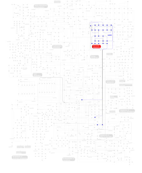
Click the image to view the interactive version of the map in iPath% proteins involved KEGG pathway ID Description 100.00  map00240
Pyrimidine metabolism This information is based on mapping of SMART genomic protein database to KEGG orthologous groups. Percentage points are related to the number of proteins with ENDO3c domain which could be assigned to a KEGG orthologous group, and not all proteins containing ENDO3c domain. Please note that proteins can be included in multiple pathways, ie. the numbers above will not always add up to 100%.
- Structure (3D structures containing this domain)
3D Structures of ENDO3c domains in PDB
PDB code Main view Title 1diz 
CRYSTAL STRUCTURE OF E. COLI 3-METHYLADENINE DNA GLYCOSYLASE (ALKA) COMPLEXED WITH DNA 1ebm 
CRYSTAL STRUCTURE OF THE HUMAN 8-OXOGUANINE GLYCOSYLASE (HOGG1) BOUND TO A SUBSTRATE OLIGONUCLEOTIDE 1fn7 
COUPLING OF DAMAGE RECOGNITION AND CATALYSIS BY A HUMAN BASE-EXCISION DNA REPAIR PROTEIN 1hu0 
CRYSTAL STRUCTURE OF AN HOGG1-DNA BOROHYDRIDE TRAPPED INTERMEDIATE COMPLEX 1kea 
STRUCTURE OF A THERMOSTABLE THYMINE-DNA GLYCOSYLASE 1kg2 
Crystal structure of the core fragment of MutY from E.coli at 1.2A resolution 1kg3 
Crystal structure of the core fragment of MutY from E.coli at 1.55A resolution 1kg4 
Crystal structure of the K142A mutant of E. coli MutY (core fragment) 1kg5 
Crystal structure of the K142Q mutant of E.coli MutY (core fragment) 1kg6 
Crystal structure of the K142R mutant of E.coli MutY (core fragment) 1kg7 
Crystal Structure of the E161A mutant of E.coli MutY (core fragment) 1ko9 
Native Structure of the Human 8-oxoguanine DNA Glycosylase hOGG1 1kqj 
Crystal Structure of a Mutant of MutY Catalytic Domain 1lwv 
Borohydride-trapped hOgg1 Intermediate Structure Co-Crystallized with 8-aminoguanine 1lww 
Borohydride-trapped hOgg1 Intermediate Structure Co-Crystallized with 8-bromoguanine 1lwy 
hOgg1 Borohydride-Trapped Intermediate without 8-oxoguanine 1m3h 
Crystal Structure of Hogg1 D268E Mutant with Product Oligonucleotide 1m3q 
Crystal Structure of hogg1 D268E Mutant with Base-Excised DNA and 8-aminoguanine 1mpg 
3-METHYLADENINE DNA GLYCOSYLASE II FROM ESCHERICHIA COLI 1mud 
CATALYTIC DOMAIN OF MUTY FROM ESCHERICHIA COLI, D138N MUTANT COMPLEXED TO ADENINE 1mun 
CATALYTIC DOMAIN OF MUTY FROM ESCHERICHIA COLI D138N MUTANT 1muy 
CATALYTIC DOMAIN OF MUTY FROM ESCHERICHIA COLI 1n39 
Structural and biochemical exploration of a critical amino acid in human 8-oxoguanine glycosylase 1n3a 
Structural and biochemical exploration of a critical amino acid in human 8-oxoguanine glycosylase 1n3c 
Structural and biochemical exploration of a critical amino acid in human 8-oxoguanine glycosylase 1orn 
Structure of a Trapped Endonuclease III-DNA Covalent Intermediate: Estranged-Guanine Complex 1orp 
Structure of a Trapped Endonuclease III-DNA Covalent Intermediate: Estranged-Adenine Complex 1p59 
Structure of a non-covalent Endonuclease III-DNA Complex 1pu6 
Crystal structure of H.pylori 3-methyladenine DNA glycosylase (MagIII) 1pu7 
Crystal structure of H.pylori 3-methyladenine DNA glycosylase (MagIII) bound to 3,9-dimethyladenine 1pu8 
Crystal structure of H.pylori 3-methyladenine DNA glycosylase (MagIII) bound to 1,N6-ethenoadenine 1pvs 
3-methyladenine Glcosylase II(AlkA) Hypoxanthine complex 1rrq 
MutY adenine glycosylase in complex with DNA containing an A:oxoG pair 1rrs 
MutY adenine glycosylase in complex with DNA containing an abasic site 1vrl 
MutY adenine glycosylase in complex with DNA and soaked adenine free base 1wef 
Catalytic Domain Of Muty From Escherichia Coli K20A Mutant 1weg 
Catalytic Domain Od Muty Form Escherichia Coli K142A Mutant 1wei 
Catalytic Domain Of Muty From Escherichia Coli K20A Mutant Complexed To Adenine 1yqk 
Human 8-oxoguanine glycosylase crosslinked with guanine containing DNA 1yql 
Catalytically inactive hOGG1 crosslinked with 7-deaza-8-azaguanine containing DNA 1yqm 
Catalytically inactive human 8-oxoguanine glycosylase crosslinked to 7-deazaguanine containing DNA 1yqr 
Catalytically inactive human 8-oxoguanine glycosylase crosslinked to oxoG containing DNA 2abk 
REFINEMENT OF THE NATIVE STRUCTURE OF ENDONUCLEASE III TO A RESOLUTION OF 1.85 ANGSTROM 2csb 
Crystal structure of Topoisomerase V from Methanopyrus kandleri (61 kDa fragment) 2csd 
Crystal structure of Topoisomerase V (61 kDa fragment) 2h56 
Crystal structure of DNA-3-methyladenine glycosidase (10174367) from Bacillus halodurans at 2.55 A resolution 2i5w 
Structure of hOGG1 crosslinked to DNA sampling a normal G adjacent to an oxoG 2jhj 
3-methyladenine dna-glycosylase from Archaeoglobus fulgidus 2jhn 
3-methyladenine dna-glycosylase from Archaeoglobus fulgidus 2nob 
Structure of catalytically inactive H270A human 8-oxoguanine glycosylase crosslinked to 8-oxoguanine DNA 2noe 
Structure of catalytically inactive G42A human 8-oxoguanine glycosylase complexed to 8-oxoguanine DNA 2nof 
Structure of Q315F human 8-oxoguanine glycosylase proximal crosslink to 8-oxoguanine DNA 2noh 
Structure of catalytically inactive Q315A human 8-oxoguanine glycosylase complexed to 8-oxoguanine DNA 2noi 
Structure of G42A human 8-oxoguanine glycosylase crosslinked to undamaged G-containing DNA 2nol 
Structure of catalytically inactive human 8-oxoguanine glycosylase distal crosslink to oxoG DNA 2noz 
Structure of Q315F human 8-oxoguanine glycosylase distal crosslink to 8-oxoguanine DNA 2xhi 
Separation-of-function mutants unravel the dual reaction mode of human 8-oxoguanine DNA glycosylase 2yg8 
Structure of an unusual 3-METHYLADENINE DNA GLYCOSYLASE II (ALKA) FROM DEINOCOCCUS RADIODURANS 2yg9 
Structure of an unusual 3-METHYLADENINE DNA GLYCOSYLASE II (ALKA) FROM DEINOCOCCUS RADIODURANS 3cvs 
Crystal Structure of an AlkA Host/Guest Complex 8oxoGuanine:Adenine Base Pair 3cvt 
Crystal Structure of an AlkA Host/Guest Complex 8oxoGuanine:Cytosine Base Pair 3cw7 
Crystal Structure of an AlkA Host/Guest Complex 8oxoGuanine:Cytosine Base Pair 3cwa 
Crystal Structure of an AlkA Host/Guest Complex 8oxoGuanine:Cytosine Base Pair 3cws 
Crystal Structure of an AlkA Host/Guest Complex 2'-fluoro-2'-deoxyinosine:Thymine Base Pair 3cwt 
Crystal Structure of an AlkA Host/Guest Complex 2'-fluoro-2'-deoxyinosine:Adenine Base Pair 3cwu 
Crystal Structure of an AlkA Host/Guest Complex 2'-fluoro-2'-deoxy-1,N6-ethenoadenine:Thymine Base Pair 3d4v 
Crystal Structure of an AlkA Host/Guest Complex N7MethylGuanine:Cytosine Base Pair 3f0z 
Crystal structure of Clostridium acetobutylicum 8-oxoguanine glycosylase/lyase in its apo-form 3f10 
Crystal structure of Clostridium Acetobutylicum 8-oxoguanine DNA glycosylase in complex with 8-oxoguanosine 3fhf 
Crystal structure of Methanocaldococcus jannaschii 8-oxoguanine DNA glycosylase (MjOgg) 3fhg 
Crystal structure of Sulfolobus solfataricus 8-oxoguanine DNA glycosylase (SsOgg) 3fsp 
MutY adenine glycosylase bound to a transition state analog (1N) paired with dG in duplexed DNA 3g0q 
Crystal Structure of MutY bound to its inhibitor DNA 3i0w 
Crystal structure of Clostridium acetobutylicum 8-oxoguanine glycosylase/lyase in complex with dsDNA containing cytosine opposite to 8-oxoG 3i0x 
Crystal structure of Clostridium acetobutylicum 8-oxoguanine glycosylase/lyase in complex with dsDNA containing adenine opposite to 8-oxoG 3ih7 
Crystal structure of catalytically active human 8-oxoguanine glycosylase distally crosslinked to guanine-containing DNA 3knt 
Crystal structure of Methanocaldococcus jannaschii 8-oxoguanine glycosylase/lyase in complex with 15mer DNA containing 8-oxoguanine 3ktu 
Structure of human 8-oxoGuanine Glycosylase 1 bound to fluorninated oxoG-containing DNA 3n0u 
Crystal structure of Tm1821, the 8-oxoguanine DNA glycosylase of Thermotoga maritima 3n5n 
Crystal structure analysis of the catalytic domain and interdomain connector of human MutY homologue 3ogd 
AlkA Undamaged DNA Complex: Interrogation of a G*:C base pair 3oh6 
AlkA Undamaged DNA Complex: Interrogation of a C:G base pair 3oh9 
AlkA Undamaged DNA Complex: Interrogation of a T:A base pair 3s6i 
Schizosaccaromyces pombe 3-methyladenine DNA glycosylase (Mag1) in complex with abasic-DNA. 4b21 
Unprecedented sculpting of DNA at abasic sites by DNA glycosylase homolog Mag2 4b22 
Unprecedented shaping of abasic sites by DNA glycosylase homolog Mag2 from Schizosaccharomyces pombe 4b23 
Unprecedented shculpting of DNA at abasic sites by DNA glycosylase homolog Mag2 4b24 
Unprecedented sculpting of DNA at abasic sites by DNA glycosylase homolog Mag2 4ejy 
Structure of MBOgg1 in complex with high affinity DNA ligand 4ejz 
Structure of MBOgg1 in complex with low affinity DNA ligand 4gfj 
Crystal structure of Topo-78, an N-terminal 78kDa fragment of topoisomerase V 4hsb 
S. pombe 3-methyladenine DNA glycosylase-like protein Mag2 bound to damaged DNA 4unf 
4UNF 4uob 
4UOB 4yoq 
4YOQ 4yph 
4YPH 4ypr 
4YPR 5an4 
5AN4 5dpk 
5DPK 5hm5 
5HM5 - Links (links to other resources describing this domain)
-
INTERPRO IPR003265 PFAM AlkA_DNA_repair PROSITE ENDONUCLEASE_III_2

