The domain within your query sequence starts at position 333 and ends at position 381; the E-value for the CBS domain shown below is 8.69e-11.
DLAVVLETAPVLTALDIFVDRRVSALPVVNESGQVVGLYSRFDVIHLAA
CBSDomain in cystathionine beta-synthase and other proteins. |
|---|
| SMART accession number: | SM00116 |
|---|---|
| Description: | Domain present in all 3 forms of cellular life. Present in two copies in inosine monophosphate dehydrogenase, of which one is disordered in the crystal structure [3]. A number of disease states are associated with CBS-containing proteins including homocystinuria, Becker's and Thomsen disease. |
| Interpro abstract (IPR000644): | CBS domains are small intracellular modules that pair together to form a stable globular domain [ (PUBMED:10200156) ]. Pairs of these domains have been termed a Bateman domain [ (PUBMED:14722609) ]. CBS domains have been shown to bind ligands with an adenosyl group such as AMP, ATP and S-AdoMet [ (PUBMED:14722619) ]. CBS domains are found attached to a wide range of other protein domains suggesting that CBS domains may play a regulatory role making proteins sensitive to adenosyl carrying ligands. The region containing the CBS domains in cystathionine-beta synthase is involved in regulation by S-AdoMet [ (PUBMED:11524006) ]. CBS domain pairs from AMPK bind AMP or ATP [ (PUBMED:14722619) ]. The CBS domains from IMPDH and the chloride channel CLC2 bind ATP [ (PUBMED:14722619) ]. |
| Family alignment: |
There are 371514 CBS domains in 203268 proteins in SMART's nrdb database.
Click on the following links for more information.
- Evolution (species in which this domain is found)
-
Taxonomic distribution of proteins containing CBS domain.
This tree includes only several representative species. The complete taxonomic breakdown of all proteins with CBS domain is also avaliable.
Click on the protein counts, or double click on taxonomic names to display all proteins containing CBS domain in the selected taxonomic class.
- Cellular role (predicted cellular role)
-
Binding / catalysis: unknown
- Literature (relevant references for this domain)
-
Primary literature is listed below; Automatically-derived, secondary literature is also avaliable.
- Bateman A
- The structure of a domain common to archaebacteria and the homocystinuria disease protein.
- Trends Biochem Sci. 1997; 22: 12-3
- Ponting CP
- CBS domains in CIC chloride channels implicated in myotonia and nephrolithiasis (kidney stones).
- J Mol Med. 1997; 75: 160-3
- Sintchak MD et al.
- Structure and mechanism of inosine monophosphate dehydrogenase in complex with the immunosuppressant mycophenolic acid.
- Cell. 1996; 85: 921-30
- Display abstract
The structure of inosine-5'-monophosphate dehydrogenase (IMPDH) in complex with IMP and mycophenolic acid (MPA) has been determined by X-ray diffraction. IMPDH plays a central role in B and T lymphocyte replication. MPA is a potent IMPDH inhibitor and the active metabolite of an immunosuppressive drug recently approved for the treatment of allograft rejection. IMPDH comprises two domains: a core domain, which is an alpha/beta barrel and contains the active site, and a flanking domain. The complex, in combination with mutagenesis and kinetic data, provides a structural basis for understanding the mechanism of IMPDH activity and indicates that MPA inhibits IMPDH by acting as a replacement for the nicotinamide portion of the nicotinamide adenine dinucleotide cofactor and a catalytic water molecule.
- Disease (disease genes where sequence variants are found in this domain)
-
SwissProt sequences and OMIM curated human diseases associated with missense mutations within the CBS domain.
Protein Disease UNKNOWN (SMART) OMIM:236200: Homocystinuria, B6-responsive and nonresponsive types - Metabolism (metabolic pathways involving proteins which contain this domain)
-

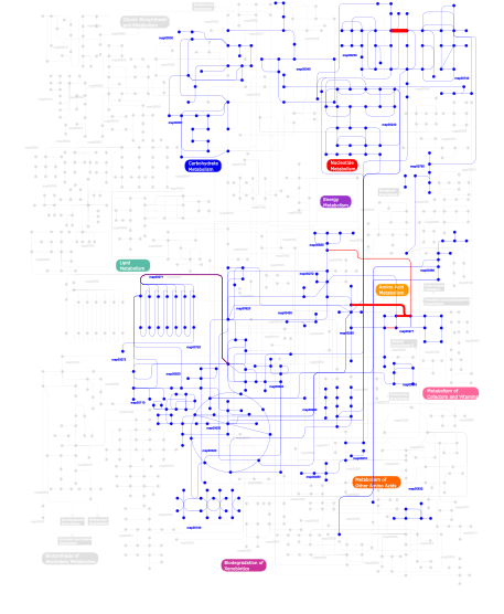
Click the image to view the interactive version of the map in iPath% proteins involved KEGG pathway ID Description 38.46  map00230
Purine metabolism 11.88 map02010 ABC transporters - General 5.94  map00450
Selenoamino acid metabolism 5.74  map00271
Methionine metabolism 4.87  map00260
Glycine, serine and threonine metabolism 4.87 map05040 Huntington's disease 3.41  map00620
Pyruvate metabolism 3.31  map00710
Carbon fixation 3.12  map00720
Reductive carboxylate cycle (CO2 fixation) 3.12  map00020
Citrate cycle (TCA cycle) 3.02  map00630
Glyoxylate and dicarboxylate metabolism 2.53  map00190
Oxidative phosphorylation 1.85  map00920
Sulfur metabolism 1.75 map04920 Adipocytokine signaling pathway 1.75 map04910 Insulin signaling pathway 1.07  map00272
Cysteine metabolism 0.88  map00051
Fructose and mannose metabolism 0.29 map02020 Two-component system - General 0.29 map03010 Ribosome 0.19  map00650
Butanoate metabolism 0.10  map00910
Nitrogen metabolism 0.10  map00040
Pentose and glucuronate interconversions 0.10  map00790
Folate biosynthesis 0.10  map00071
Fatty acid metabolism 0.10  map00632
Benzoate degradation via CoA ligation 0.10  map00740
Riboflavin metabolism 0.10 map03020 RNA polymerase 0.10 map02060 Phosphotransferase system (PTS) 0.10  map00280
Valine, leucine and isoleucine degradation 0.10  map00240
Pyrimidine metabolism 0.10  map00072
Synthesis and degradation of ketone bodies 0.10  map00310
Lysine degradation 0.10 map01051 Biosynthesis of ansamycins 0.10  map00500
Starch and sucrose metabolism 0.10  map00380
Tryptophan metabolism 0.10  map00480
Glutathione metabolism 0.10  map00640
Propanoate metabolism This information is based on mapping of SMART genomic protein database to KEGG orthologous groups. Percentage points are related to the number of proteins with CBS domain which could be assigned to a KEGG orthologous group, and not all proteins containing CBS domain. Please note that proteins can be included in multiple pathways, ie. the numbers above will not always add up to 100%.
- Structure (3D structures containing this domain)
3D Structures of CBS domains in PDB
PDB code Main view Title 1ak5 
INOSINE MONOPHOSPHATE DEHYDROGENASE (IMPDH) FROM TRITRICHOMONAS FOETUS 1b3o 
TERNARY COMPLEX OF HUMAN TYPE-II INOSINE MONOPHOSPHATE DEHYDROGENASE WITH 6-CL-IMP AND SELENAZOLE ADENINE DINUCLEOTIDE 1jcn 
BINARY COMPLEX OF HUMAN TYPE-I INOSINE MONOPHOSPHATE DEHYDROGENASE WITH 6-CL-IMP 1jr1 
Crystal structure of Inosine Monophosphate Dehydrogenase in complex with Mycophenolic Acid 1me7 
Inosine Monophosphate Dehydrogenase (IMPDH) From Tritrichomonas Foetus with RVP and MOA bound 1me8 
Inosine Monophosphate Dehydrogenase (IMPDH) From Tritrichomonas Foetus with RVP bound 1me9 
Inosine Monophosphate Dehydrogenase (IMPDH) From Tritrichomonas Foetus with IMP bound 1meh 
Inosine Monophosphate Dehydrogenase (IMPDH) From Tritrichomonas Foetus with IMP and MOA bound 1mei 
Inosine Monophosphate Dehydrogenase (IMPDH) From Tritrichomonas Foetus with XMP and mycophenolic acid bound 1mew 
Inosine Monophosphate Dehydrogenase (IMPDH) From Tritrichomonas Foetus with XMP and NAD bound 1nf7 
Ternary complex of the human type II Inosine Monophosphate Dedhydrogenase with Ribavirin Monophosphate and C2-Mycophenolic Adenine Dinucleotide 1nfb 
Ternary complex of the human type II Inosine Monophosphate Dedhydrogenase with 6Cl-IMP and NAD 1o50 
Crystal structure of a cbs domain-containing protein (tm0935) from thermotoga maritima at 1.87 A resolution 1pbj 
CBS domain protein 1pvm 
Crystal Structure of a Conserved CBS Domain Protein TA0289 of Unknown Function from Thermoplasma acidophilum 1vr9 
CRYSTAL STRUCTURE OF A CBS DOMAIN PAIR/ACT DOMAIN PROTEIN (TM0892) FROM THERMOTOGA MARITIMA AT 1.70 A RESOLUTION 1vrd 
Crystal structure of Inosine-5'-monophosphate dehydrogenase (TM1347) from THERMOTOGA MARITIMA at 2.18 A resolution 1xkf 
Crystal structure of Hypoxic Response Protein I (HRPI) with two coordinated zinc ions 1y5h 
Crystal structure of truncated Se-Met Hypoxic Response Protein I (HRPI) 1zfj 
INOSINE MONOPHOSPHATE DEHYDROGENASE (IMPDH; EC 1.1.1.205) FROM STREPTOCOCCUS PYOGENES 2cu0 
Crystal structure of inosine-5'-monophosphate dehydrogenase from Pyrococcus horikoshii OT3 2d4z 
Crystal structure of the cytoplasmic domain of the chloride channel ClC-0 2ef7 
Crystal structure of ST2348, a hypothetical protein with CBS domains from Sulfolobus tokodaii strain7 2emq 
Hypothetical Conserved Protein (GK1048) from Geobacillus Kaustophilus 2j9l 
Cytoplasmic Domain of the Human Chloride Transporter ClC-5 in complex with ATP 2ja3 
Cytoplasmic Domain of the Human Chloride Transporter ClC-5 in complex with ADP 2nyc 
Crystal structure of the Bateman2 domain of yeast Snf4 2nye 
Crystal structure of the Bateman2 domain of yeast Snf4 2o16 
Crystal structure of a putative acetoin utilization protein (AcuB) from Vibrio cholerae 2oox 
Crystal structure of the adenylate sensor from AMP-activated protein kinase complexed with AMP 2ooy 
Crystal structure of the adenylate sensor from AMP-activated protein kinase complexed with ATP 2oux 
Crystal structure of the soluble part of a magnesium transporter 2p9m 
Crystal structure of conserved hypothetical protein MJ0922 from Methanocaldococcus jannaschii DSM 2661 2pfi 
Crystal structure of the cytoplasmic domain of the human chloride channel ClC-Ka 2qh1 
Structure of TA289, a CBS-rubredoxin-like protein, in its Fe+2-bound state 2qlv 
Crystal structure of the heterotrimer core of the S. cerevisiae AMPK homolog SNF1 2qr1 
Crystal structure of the adenylate sensor from AMP-activated protein kinase in complex with ADP 2qrc 
Crystal structure of the adenylate sensor from AMP-activated protein kinase in complex with ADP and AMP 2qrd 
Crystal Structure of the Adenylate Sensor from AMP-activated Protein Kinase in complex with ADP and ATP 2qre 
Crystal structure of the adenylate sensor from AMP-activated protein kinase in complex with 5-aminoimidazole-4-carboxamide 1-beta-D-ribofuranotide (ZMP) 2rc3 
Crystal structure of CBS domain, NE2398 2rif 
CBS domain protein PAE2072 from Pyrobaculum aerophilum complexed with AMP 2rih 
CBS domain protein PAE2072 from Pyrobaculum aerophilum 2uv4 
Crystal Structure of a CBS domain pair from the regulatory gamma1 subunit of human AMPK in complex with AMP 2uv5 
Crystal Structure of a CBS domain pair from the regulatory gamma1 subunit of human AMPK in complex with AMP 2uv6 
Crystal Structure of a CBS domain pair from the regulatory gamma1 subunit of human AMPK in complex with AMP 2uv7 
Crystal Structure of a CBS domain pair from the regulatory gamma1 subunit of human AMPK in complex with AMP 2v8q 
Crystal structure of the regulatory fragment of mammalian AMPK in complexes with AMP 2v92 
Crystal structure of the regulatory fragment of mammalian AMPK in complexes with ATP-AMP 2v9j 
Crystal structure of the regulatory fragment of mammalian AMPK in complexes with Mg.ATP-AMP 2y8l 
Structure of an active form of mammalian AMPK in complex with two ADP 2y8q 
Structure of the regulatory fragment of mammalian AMPK in complex with one ADP 2ya3 
STRUCTURE OF THE REGULATORY FRAGMENT OF MAMMALIAN AMPK IN COMPLEX WITH COUMARIN ADP 2yvx 
Crystal structure of magnesium transporter MgtE 2yvy 
Crystal structure of magnesium transporter MgtE cytosolic domain, Mg2+ bound form 2yvz 
Crystal structure of magnesium transporter MgtE cytosolic domain, Mg2+-free form 2yzi 
Crystal structure of uncharacterized conserved protein from Pyrococcus horikoshii 2yzq 
Crystal structure of uncharacterized conserved protein from Pyrococcus horikoshii 2zy9 
Improved crystal structure of magnesium transporter MgtE 3ddj 
Crystal structure of a cbs domain-containing protein in complex with amp (sso3205) from sulfolobus solfataricus at 1.80 A resolution 3fhm 
Crystal structure of the CBS-domain containing protein ATU1752 from Agrobacterium tumefaciens 3fv6 
Crystal Structure of the CBS domains from the Bacillus subtilis CcpN repressor 3fwr 
Crystal Structure of the CBS domains from the Bacillus subtilis CcpN repressor complexed with ADP 3fws 
Crystal Structure of the CBS domains from the Bacillus subtilis CcpN repressor complexed with AppNp, phosphate and magnesium ions 3ghd 
Crystal structure of a cystathionine beta-synthase domain protein fused to a Zn-ribbon-like domain 3hf7 
The Crystal Structure of a CBS-domain Pair with Bound AMP from Klebsiella pneumoniae to 2.75A 3i8n 
A domain of a conserved functionally known protein from Vibrio parahaemolyticus RIMD 2210633. 3jtf 
The CBS Domain Pair Structure of a magnesium and cobalt efflux protein from Bordetella parapertussis in complex with AMP 3k6e 
Crystal structure of cbs domain protein from streptococcus pneumoniae tigr4 3kh5 
Crystal Structure of Protein MJ1225 from Methanocaldococcus jannaschii, a putative archaeal homolog of g-AMPK. 3kpb 
Crystal Structure of the CBS domain pair of protein MJ0100 in complex with 5 -methylthioadenosine and S-adenosyl-L-methionine. 3kpc 
Crystal Structure of the CBS domain pair of protein MJ0100 in complex with 5 -methylthioadenosine and S-adenosyl-L-methionine 3kpd 
Crystal Structure of the CBS domain pair of protein MJ0100 in complex with 5 -methylthioadenosine and S-adenosyl-L-methionine. 3kxr 
Structure of the cystathionine beta-synthase pair domain of the putative Mg2+ transporter SO5017 from Shewanella oneidensis MR-1. 3l2b 
Crystal structure of the CBS and DRTGG domains of the regulatory region of Clostridium perfringens pyrophosphatase complexed with activator, diadenosine tetraphosphate 3l31 
Crystal structure of the CBS and DRTGG domains of the regulatory region of Clostridium perfringens pyrophosphatase complexed with the inhibitor, AMP 3lfr 
The Crystal Structure of a CBS Domain from a Putative Metal Ion Transporter Bound to AMP from Pseudomonas syringae to 1.55A 3lfz 
Crystal Structure of Protein MJ1225 from Methanocaldococcus jannaschii, a putative archaeal homolog of g-AMPK. 3lqn 
Crystal Structure of CBS Domain-containing Protein of Unknown Function from Bacillus anthracis str. Ames Ancestor 3lv9 
Crystal structure of CBS domain of a putative transporter from Clostridium difficile 630 3nqr 
A putative CBS domain-containing protein from Salmonella typhimurium LT2 3oco 
The crystal structure of a Hemolysin-like protein containing CBS domain of Oenococcus oeni PSU 3oi8 
The crystal structure of functionally unknown conserved protein domain from Neisseria meningitidis MC58 3pc2 
Full length structure of cystathionine beta-synthase from Drosophila 3pc3 
Full length structure of cystathionine beta-synthase from Drosophila in complex with aminoacrylate 3pc4 
Full length structure of cystathionine beta-synthase from Drosophila in complex with serine 3sl7 
Crystal structure of CBS-pair protein, CBSX2 from Arabidopsis thaliana 3t4n 
Structure of the regulatory fragment of Saccharomyces cerevisiae AMPK in complex with ADP 3tdh 
Structure of the regulatory fragment of sccharomyces cerevisiae AMPK in complex with AMP 3te5 
structure of the regulatory fragment of sacchromyces cerevisiae ampk in complex with NADH 3tsb 
Crystal Structure of Inosine-5'-monophosphate Dehydrogenase from Bacillus anthracis str. Ames 3tsd 
Crystal Structure of Inosine-5'-monophosphate Dehydrogenase from Bacillus anthracis str. Ames complexed with XMP 3usb 
Crystal Structure of Bacillus anthracis Inosine Monophosphate Dehydrogenase in the complex with IMP 3zfh 
Crystal structure of Pseudomonas aeruginosa inosine 5'-monophosphate dehydrogenase 4af0 
Crystal structure of cryptococcal inosine monophosphate dehydrogenase 4avf 
Crystal structure of Pseudomonas aeruginosa inosine 5'-monophosphate dehydrogenase 4cfe 
Structure of full length human AMPK in complex with a small molecule activator, a benzimidazole derivative (991) 4cff 
Structure of full length human AMPK in complex with a small molecule activator, a thienopyridone derivative (A-769662) 4cfh 
Structure of an active form of mammalian AMPK 4coo 
Crystal structure of human cystathionine beta-synthase (delta516-525) at 2.0 angstrom resolution 4dqw 
Crystal Structure Analysis of PA3770 4eag 
Co-crystal structure of an chimeric AMPK core with ATP 4eai 
Co-crystal structure of an AMPK core with AMP 4eaj 
Co-crystal of AMPK core with AMP soaked with ATP 4eak 
Co-crystal structure of an AMPK core with ATP 4eal 
Co-crystal of AMPK core with ATP soaked with AMP 4esy 
Crystal Structure of the CBS Domain of CBS Domain Containing Membrane Protein from Sphaerobacter thermophilus 4fry 
The structure of a putative signal-transduction protein with CBS domains from Burkholderia ambifaria MC40-6 4fxs 
Inosine 5'-monophosphate dehydrogenase from Vibrio cholerae complexed with IMP and mycophenolic acid 4gqv 
Crystal structure of CBS-pair protein, CBSX1 from Arabidopsis thaliana 4gqw 
Crystal structure of CBS-pair protein, CBSX1 (loop deletion) from Arabidopsis thaliana 4gqy 
Crystal structure of CBSX2 in complex with AMP 4hg0 
Crystal Structure of magnesium and cobalt efflux protein CorC, Northeast Structural Genomics Consortium (NESG) Target ER40 4l0d 
Crystal structure of delta516-525 human cystathionine beta-synthase containing C-terminal 6xHis-tag 4l27 
Crystal structure of delta1-39 and delta516-525 human cystathionine beta-synthase D444N mutant containing C-terminal 6xHis tag 4l28 
Crystal structure of delta516-525 human cystathionine beta-synthase D444N mutant containing C-terminal 6xHis tag 4l3v 
Crystal structure of delta516-525 human cystathionine beta-synthase 4noc 
The crystal structure of a CBS Domain-containing Protein of Unknown Function from Kribbella flavida DSM 17836. 4o9k 
Crystal structure of the CBS pair of a putative D-arabinose 5-phosphate isomerase from Methylococcus capsulatus in complex with CMP-Kdo 4pcu 
4PCU 4qfg 
4QFG 4qfr 
4QFR 4qfs 
4QFS 4rer 
4RER 4rew 
4REW 4uuu 
4UUU 4z87 
4Z87 4zhx 
4ZHX 5ahn 
5AHN 5awe 
5AWE 5ezv 
5EZV 5iip 
5IIP 5kq5 
5KQ5 5ks7 
5KS7 - Links (links to other resources describing this domain)
-
INTERPRO IPR000644

