The domain within your query sequence starts at position 525 and ends at position 589; the E-value for the DSRM domain shown below is 2.73e-21.
GKNPVMELNEKRRGLKYELISETGGSHDKRFVMEVEVDGQKFQGAGSNKKVAKAYAALAA
LEKLF
DSRMDouble-stranded RNA binding motif |
|---|
| SMART accession number: | SM00358 |
|---|---|
| Description: | - |
| Interpro abstract (IPR014720): | In contrast to other RNA-binding domains, the about 65 amino acids long dsRBD domain [ (PUBMED:1438302) (PUBMED:8036511) (PUBMED:7972084) ] has been found in a number of proteins that specifically recognise double-stranded RNAs. The dsRBD domain is also known as DSRM (Double-Stranded RNA-binding Motif). dsRBD proteins are mainly involved in posttranscriptional gene regulation, for example by preventing the expression of proteins or by mediating RNAs localization. This domain is also found in RNA editing proteins. Interaction of the dsRBD with RNA is unlikely to involve the recognition of specific sequences [ (PUBMED:1357546) (PUBMED:7527340) (PUBMED:8127710) ]. Nevertheless, multiple dsRBDs may be able to act in combination to recognise the secondary structure of specific RNAs (i.e. Staufen) [ (PUBMED:1438302) ]. NMR analysis of the third dsRBD of Drosophila Staufen have revealed an alpha-beta-beta-beta-alpha structure [ (PUBMED:7628456) ]. |
| Family alignment: |
There are 49577 DSRM domains in 36103 proteins in SMART's nrdb database.
Click on the following links for more information.
- Evolution (species in which this domain is found)
-
Taxonomic distribution of proteins containing DSRM domain.
This tree includes only several representative species. The complete taxonomic breakdown of all proteins with DSRM domain is also avaliable.
Click on the protein counts, or double click on taxonomic names to display all proteins containing DSRM domain in the selected taxonomic class.
- Cellular role (predicted cellular role)
-
Binding / catalysis: RNA binding
- Literature (relevant references for this domain)
-
Primary literature is listed below; Automatically-derived, secondary literature is also avaliable.
- Mian IS
- Comparative sequence analysis of ribonucleases HII, III, II PH and D.
- Nucleic Acids Res. 1997; 25: 3187-95
- Display abstract
Escherichia coli ribonucleases (RNases) HII, III, II, PH and D have been used to characterise new and known viral, bacterial, archaeal and eucaryotic sequences similar to these endo- (HII and III) and exoribonucleases (II, PH and D). Statistical models, hidden Markov models (HMMs), were created for the RNase HII, III, II and PH and D families as well as a double-stranded RNA binding domain present in RNase III. Results suggest that the RNase D family, which includes Werner syndrome protein and the 100 kDa antigenic component of the human polymyositis scleroderma (PMSCL) autoantigen, is a 3'-->5' exoribonuclease structurally and functionally related to the 3'-->5' exodeoxyribonuclease domain of DNA polymerases. Polynucleotide phosphorylases and the RNase PH family, which includes the 75 kDa PMSCL autoantigen, possess a common domain suggesting similar structures and mechanisms of action for these 3'-->5' phosphorolytic enzymes. Examination of HMM-generated multiple sequences alignments for each family suggest amino acids that may be important for their structure, substrate binding and/or catalysis.
- Zamore PD, Williamson JR, Lehmann R
- The Pumilio protein binds RNA through a conserved domain that defines a new class of RNA-binding proteins.
- RNA. 1997; 3: 1421-33
- Display abstract
Translation of hunchback(mat) (hb[mat]) mRNA must be repressed in the posterior of the pre-blastoderm Drosophila embryo to permit formation of abdominal segments. This translational repression requires two copies of the Nanos Response Element (NRE), a 16-nt sequence in the hb[mat] 3' untranslated region. Translational repression also requires the action of two proteins: Pumilio (PUM), a sequence-specific RNA-binding protein; and Nanos, a protein that determines the location of repression. Binding of PUM to the NRE is thought to target hb(mat) mRNA for repression. Here, we show the RNA-binding domain of PUM to be an evolutionarily conserved, 334-amino acid region at the carboxy-terminus of the approximately 158-kDa PUM protein. This contiguous region of PUM retains the RNA-binding specificity of full-length PUM protein. Proteins with sequences homologous to the PUM RNA-binding domain are found in animals, plants, and fungi. The high degree of sequence conservation of the PUM RNA-binding domain in other far-flung species suggests that the domain is an ancient protein motif, and we show that conservation of sequence reflects conservation of function: that is, the homologous region from a human protein binds RNA with sequence specificity related to but distinct from Drosophila PUM.
- Burd CG, Dreyfuss G
- Conserved structures and diversity of functions of RNA-binding proteins.
- Science. 1994; 265: 615-21
- Display abstract
In eukaryotic cells, a multitude of RNA-binding proteins play key roles in the posttranscriptional regulation of gene expression. Characterization of these proteins has led to the identification of several RNA-binding motifs, and recent experiments have begun to illustrate how several of them bind RNA. The significance of these interactions is reflected in the recent discoveries that several human and other vertebrate genetic disorders are caused by aberrant expression of RNA-binding proteins. The major RNA-binding motifs are described and examples of how they may function are given.
- StJohnston D, Brown NH, Gall JG, Jantsch M
- A conserved double-stranded RNA-binding domain.
- Proc Natl Acad Sci U S A. 1992; 89: 10979-83
- Display abstract
We have identified a double-stranded (ds)RNA-binding domain in each of two proteins: the product of the Drosophila gene staufen, which is required for the localization of maternal mRNAs, and a protein of unknown function, Xlrbpa, from Xenopus. The amino acid sequences of the binding domains are similar to each other and to additional domains in each protein. Database searches identified similar domains in several other proteins known or thought to bind dsRNA, including human dsRNA-activated inhibitor (DAI), human trans-activating region (TAR)-binding protein, and Escherichia coli RNase III. By analyzing in detail one domain in staufen and one in Xlrbpa, we delimited the minimal region that binds dsRNA. On the basis of the binding studies and computer analysis, we have derived a consensus sequence that defines a 65- to 68-amino acid dsRNA-binding domain.
- Metabolism (metabolic pathways involving proteins which contain this domain)
-

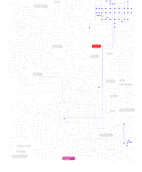
Click the image to view the interactive version of the map in iPath% proteins involved KEGG pathway ID Description 70.00  map00791
Atrazine degradation 20.00  map00230
Purine metabolism 10.00 map03010 Ribosome This information is based on mapping of SMART genomic protein database to KEGG orthologous groups. Percentage points are related to the number of proteins with DSRM domain which could be assigned to a KEGG orthologous group, and not all proteins containing DSRM domain. Please note that proteins can be included in multiple pathways, ie. the numbers above will not always add up to 100%.
- Structure (3D structures containing this domain)
3D Structures of DSRM domains in PDB
PDB code Main view Title 1di2 
CRYSTAL STRUCTURE OF A DSRNA-BINDING DOMAIN COMPLEXED WITH DSRNA: MOLECULAR BASIS OF DOUBLE-STRANDED RNA-PROTEIN INTERACTIONS 1ekz 
NMR STRUCTURE OF THE COMPLEX BETWEEN THE THIRD DSRBD FROM DROSOPHILA STAUFEN AND A RNA HAIRPIN 1o0w 
Crystal structure of Ribonuclease III (TM1102) from Thermotoga maritima at 2.0 A resolution 1qu6 
STRUCTURE OF THE DOUBLE-STRANDED RNA-BINDING DOMAIN OF THE PROTEIN KINASE PKR REVEALS THE MOLECULAR BASIS OF ITS DSRNA-MEDIATED ACTIVATION 1rc7 
Crystal structure of RNase III Mutant E110K from Aquifex Aeolicus complexed with ds-RNA at 2.15 Angstrom Resolution 1stu 
DOUBLE STRANDED RNA BINDING DOMAIN 1t4l 
Solution structure of double-stranded RNA binding domain of S. cerevisiae RNase III (Rnt1p) in complex with the 5' terminal RNA hairpin of snR47 precursor 1t4n 
Solution structure of Rnt1p dsRBD 1t4o 
Crystal structure of rnt1p dsRBD 1uhz 
Solution structure of dsRNA binding domain in Staufen homolog 2 1uil 
Double-stranded RNA-binding motif of Hypothetical protein BAB28848 1whn 
Solution structure of the dsRBD from hypothetical protein BAB26260 1whq 
Solution structure of the N-terminal dsRBD from hypothetical protein BAB28848 1x47 
Solution structure of DSRM domain in DGCR8 protein 1x48 
Solution structure of the second DSRM domain in Interferon-induced, double-stranded RNA-activated protein kinase 1x49 
Solution structure of the first DSRM domain in Interferon-induced, double-stranded RNA-activated protein kinase 1yyk 
Crystal structure of RNase III from Aquifex Aeolicus complexed with double-stranded RNA at 2.5-angstrom resolution 1yyo 
Crystal structure of RNase III mutant E110K from Aquifex aeolicus complexed with double-stranded RNA at 2.9-Angstrom Resolution 1yyw 
Crystal structure of RNase III from Aquifex aeolicus complexed with double stranded RNA at 2.8-Angstrom Resolution 1yz9 
Crystal structure of RNase III mutant E110Q from Aquifex aeolicus complexed with double stranded RNA at 2.1-Angstrom Resolution 2a11 
Crystal Structure of Nuclease Domain of Ribonuclase III from Mycobacterium Tuberculosis 2b7t 
Structure of ADAR2 dsRBM1 2b7v 
Structure of ADAR2 dsRBM2 2cpn 
Solution structure of the second dsRBD of TAR RNA-binding protein 2 2dix 
Solution structure of the DSRM domain of Protein activator of the interferon-induced protein kinase 2dmy 
Solution structure of DSRM domain in Spermatid perinuclear RNA-bind protein 2ez6 
Crystal structure of Aquifex aeolicus RNase III (D44N) complexed with product of double-stranded RNA processing 2khx 
Drosha double-stranded RNA binding motif 2l2k 
Solution NMR structure of the R/G STEM LOOP RNA-ADAR2 DSRBM2 Complex 2l2m 
Solution structure of the second dsRBD of HYL1 2l2n 
Backbone 1H, 13C, and 15N Chemical Shift Assignments for the first dsRBD of protein HYL1 2l33 
Solution NMR Structure of DRBM 2 domain of Interleukin enhancer-binding factor 3 from Homo sapiens, Northeast Structural Genomics Consortium Target HR4527E 2l3c 
Solution structure of ADAR2 dsRBM1 bound to LSL RNA 2l3j 
The solution structure of the ADAR2 dsRBM-RNA complex reveals a sequence-specific read out of the minor groove 2lbs 
Solution structure of double-stranded RNA binding domain of S. cerevisiae RNase III (Rnt1p) in complex with AAGU tetraloop hairpin 2ljh 
NMR structure of Double-stranded RNA-specific editase Adar 2lrs 
The second dsRBD domain from A. thaliana DICER-LIKE 1 2ltr 
Solution structure of RDE-4(32-136) 2lts 
Solution structure of RDE-4(150-235) 2lup 
RDC refined solution structure of double-stranded RNA binding domain of S. cerevisiae RNase III (rnt1p) in complex with the terminal RNA hairpin of snr47 precursor 2luq 
Solution structure of double-stranded RNA binding domain of S.cerevisiae RNase III (rnt1p) 2mdr 
Solution structure of the third double-stranded RNA-binding domain (dsRBD3) of human adenosine-deaminase ADAR1 2n3f 
2N3F 2n3g 
2N3G 2n3h 
2N3H 2nue 
Crystal structure of RNase III from Aquifex aeolicus complexed with ds-RNA at 2.9-Angstrom Resolution 2nuf 
Crystal structure of RNase III from Aquifex aeolicus complexed with ds-RNA at 2.5-Angstrom Resolution 2nug 
Crystal structure of RNase III from Aquifex aeolicus complexed with ds-RNA at 1.7-Angstrom Resolution 2rs6 
Solution structure of the N-terminal dsRBD from RNA helicase A 2rs7 
Solution structure of the second dsRBD from RNA helicase A 2yt4 
Crystal structure of human DGCR8 core 3adg 
Structure of Arabidopsis HYL1 and its molecular implications for miRNA processing 3adi 
Structure of Arabidopsis HYL1 and its molecular implications for miRNA processing 3adj 
Structure of Arabidopsis HYL1 and its molecular implications for miRNA processing 3adl 
Structure of TRBP2 and its molecule implications for miRNA processing 3c4b 
Structure of RNaseIIIb and dsRNA binding domains of mouse Dicer 3c4t 
Structure of RNaseIIIb and dsRNA binding domains of mouse Dicer 3htx 
Crystal structure of small RNA methyltransferase HEN1 3j6b 
3J6B 3llh 
Crystal structure of the first dsRBD of TAR RNA-binding protein 2 3n3w 
2.2 Angstrom Resolution Crystal Structure of Nuclease Domain of Ribonuclase III (rnc) from Campylobacter jejuni 3p1x 
Crystal structure of DRBM 2 domain of Interleukin enhancer-binding factor 3 from Homo sapiens, Northeast Structural Genomics Consortium Target HR4527E 3rv0 
Crystal structure of K. polysporus Dcr1 without the C-terminal dsRBD 3vyx 
Structural insights into RISC assembly facilitated by dsRNA binding domains of human RNA helicase (DHX9) 3vyy 
Structural insights into RISC assembly facilitated by dsRNA binding domains of human RNA helicase A (DHX9) 4m2z 
Crystal structure of RNASE III complexed with double-stranded RNA and CMP (TYPE II CLEAVAGE) 4m30 
Crystal structure of RNASE III complexed with double-stranded RNA AND AMP (TYPE II CLEAVAGE) 4oog 
Crystal structure of yeast RNase III (Rnt1p) complexed with the product of dsRNA processing 4wft 
4WFT 4x8w 
4X8W 5aor 
5AOR 5b16 
5B16 5dv7 
5DV7 - Links (links to other resources describing this domain)
-
INTERPRO IPR014720 PFAM dsrm

