The domain within your query sequence starts at position 155 and ends at position 221; the E-value for the FYVE domain shown below is 1.81e-31.
ERAPDWVDAEECHRCRVQFGVVTRKHHCRACGQIFCGKCSSKYSTIPKFGIEKEVRVCEP
CYEQLNK
FYVEProtein present in Fab1, YOTB, Vac1, and EEA1 |
|---|
| SMART accession number: | SM00064 |
|---|---|
| Description: | The FYVE zinc finger is named after four proteins where it was first found: Fab1, YOTB/ZK632.12, Vac1, and EEA1. The FYVE finger has been shown to bind two Zn2+ ions. The FYVE finger has eight potential zinc coordinating cysteine positions. The FYVE finger is structurally related to the PHD finger and the RING finger. Many members of this family also include two histidines in a motif R+HHC+XCG, where + represents a charged residue and X any residue. The FYVE finger functions in the membrane recruitment of cytosolic proteins by binding to phosphatidylinositol 3-phosphate (PI3P), which is prominent on endosomes. The R+HHC+XCG motif is critical for PI3P binding. |
| Interpro abstract (IPR000306): | Zinc finger (Znf) domains are relatively small protein motifs which contain multiple finger-like protrusions that make tandem contacts with their target molecule. Some of these domains bind zinc, but many do not; instead binding other metals such as iron, or no metal at all. For example, some family members form salt bridges to stabilise the finger-like folds. They were first identified as a DNA-binding motif in transcription factor TFIIIA from Xenopus laevis (African clawed frog), however they are now recognised to bind DNA, RNA, protein and/or lipid substrates [ (PUBMED:10529348) (PUBMED:15963892) (PUBMED:15718139) (PUBMED:17210253) (PUBMED:12665246) ]. Their binding properties depend on the amino acid sequence of the finger domains and of the linker between fingers, as well as on the higher-order structures and the number of fingers. Znf domains are often found in clusters, where fingers can have different binding specificities. There are many superfamilies of Znf motifs, varying in both sequence and structure. They display considerable versatility in binding modes, even between members of the same class (e.g. some bind DNA, others protein), suggesting that Znf motifs are stable scaffolds that have evolved specialised functions. For example, Znf-containing proteins function in gene transcription, translation, mRNA trafficking, cytoskeleton organisation, epithelial development, cell adhesion, protein folding, chromatin remodelling and zinc sensing, to name but a few [ (PUBMED:11179890) ]. Zinc-binding motifs are stable structures, and they rarely undergo conformational changes upon binding their target. The FYVE zinc finger is named after four proteins that it has been found in: Fab1, YOTB/ZK632.12, Vac1, and EEA1. The FYVE finger has been shown to bind two zinc ions [ (PUBMED:8798641) ]. The FYVE finger has eight potential zinc coordinating cysteine positions. Many members of this family also include two histidines in a motif R+HHC+XCG, where + represents a charged residue and X any residue. FYVE-type domains are divided into two known classes: FYVE domains that specifically bind to phosphatidylinositol 3-phosphate in lipid bilayers and FYVE-related domains of undetermined function [ (PUBMED:15576038) ]. Those that bind to phosphatidylinositol 3-phosphate are often found in proteins targeted to lipid membranes that are involved in regulating membrane traffic [ (PUBMED:11456498) (PUBMED:11739631) (PUBMED:11509568) ]. Most FYVE domains target proteins to endosomes by binding specifically to phosphatidylinositol-3-phosphate at the membrane surface. By contrast, the CARP2 FYVE-like domain is not optimized to bind to phosphoinositides or insert into lipid bilayers. FYVE domains are distinguished from other zinc fingers by three signature sequences: an N-terminal WxxD motif, a basic R(R/K)HHCR patch, and a C-terminal RVC motif. |
| GO function: | metal ion binding (GO:0046872) |
| Family alignment: |
There are 26595 FYVE domains in 25311 proteins in SMART's nrdb database.
Click on the following links for more information.
- Evolution (species in which this domain is found)
- Cellular role (predicted cellular role)
- Literature (relevant references for this domain)
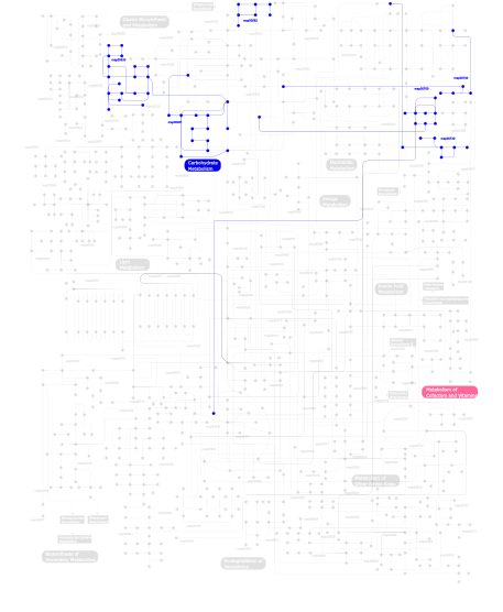
- Metabolism (metabolic pathways involving proteins which contain this domain)
- Structure (3D structures containing this domain)
- Links (links to other resources describing this domain)


