The domain within your query sequence starts at position 847 and ends at position 1049; the E-value for the MUTSac domain shown below is 9.7e-122.
RVMIITGPNMGGKSSYIKQVALVTIMAQIGSYVPAEEATIGIVDGIFTRMGAADNIYKGR
STFMEELTDTAEIIRRASPQSLVILDELGRGTSTHDGIAIAYATLEYFIRDVKSLTLFVT
HYPPVCELEKCYPEQVGNYHMGFLVNEDESKQDSGDMEQMPDSVTFLYQITRGIAARSYG
LNVAKLADVPREVLQKAAHKSKE
MUTSacATPase domain of DNA mismatch repair MUTS family |
|---|
| SMART accession number: | SM00534 |
|---|---|
| Description: | - |
| Interpro abstract (IPR000432): | Mismatch repair contributes to the overall fidelity of DNA replication and is essential for combating the adverse effects of damage to the genome. It involves the correction of mismatched base pairs that have been missed by the proofreading element of the DNA polymerase complex. The post-replicative Mismatch Repair System (MMRS) of Escherichia coli involves MutS (Mutator S), MutL and MutH proteins, and acts to correct point mutations or small insertion/deletion loops produced during DNA replication [ (PUBMED:17919654) ]. MutS and MutL are involved in preventing recombination between partially homologous DNA sequences. The assembly of MMRS is initiated by MutS, which recognises and binds to mispaired nucleotides and allows further action of MutL and MutH to eliminate a portion of newly synthesized DNA strand containing the mispaired base [ (PUBMED:17599803) ]. MutS can also collaborate with methyltransferases in the repair of O(6)-methylguanine damage, which would otherwise pair with thymine during replication to create an O(6)mG:T mismatch [ (PUBMED:17951114) ]. MutS exists as a dimer, where the two monomers have different conformations and form a heterodimer at the structural level [ (PUBMED:17426027) ]. Only one monomer recognises the mismatch specifically and has ADP bound. Non-specific major groove DNA-binding domains from both monomers embrace the DNA in a clamp-like structure. Mismatch binding induces ATP uptake and a conformational change in the MutS protein, resulting in a clamp that translocates on DNA. MutS is a modular protein with a complex structure [ (PUBMED:11048711) ], and is composed of:
The MutS family of proteins is named after the Salmonella typhimurium MutS protein involved in mismatch repair. Homologues of MutS have been found in many species including eukaryotes (MSH 1, 2, 3, 4, 5, and 6 proteins), archaea and bacteria, and together these proteins have been grouped into the MutS family. Although many of these proteins have similar activities to the E. coli MutS, there is significant diversity of function among the MutS family members. Human MSH has been implicated in non-polyposis colorectal carcinoma (HNPCC) and is a mismatch binding protein [ (PUBMED:8036718) ].This diversity is even seen within species, where many species encode multiple MutS homologues with distinct functions [ (PUBMED:9722651) ]. Inter-species homologues may have arisen through frequent ancient horizontal gene transfer of MutS (and MutL) from bacteria to archaea and eukaryotes via endosymbiotic ancestors of mitochondria and chloroplasts [ (PUBMED:17965091) ]. This entry represents the C-terminal domain found in proteins in the MutS family of DNA mismatch repair proteins. The C-terminal region of MutS is comprised of the ATPase domain and the HTH (helix-turn-helix) domain, the latter being involved in dimer contacts. Yeast MSH3 [ (PUBMED:8510668) ], bacterial proteins involved in DNA mismatch repair, and the predicted protein product of the Rep-3 gene of mouse share extensive sequence similarity. Human MSH has been implicated in non-polyposis colorectal carcinoma (HNPCC) and is a mismatch binding protein. |
| GO process: | mismatch repair (GO:0006298) |
| GO function: | mismatched DNA binding (GO:0030983), ATP binding (GO:0005524) |
| Family alignment: |
There are 42358 MUTSac domains in 42333 proteins in SMART's nrdb database.
Click on the following links for more information.
- Evolution (species in which this domain is found)
- Literature (relevant references for this domain)
- Disease (disease genes where sequence variants are found in this domain)
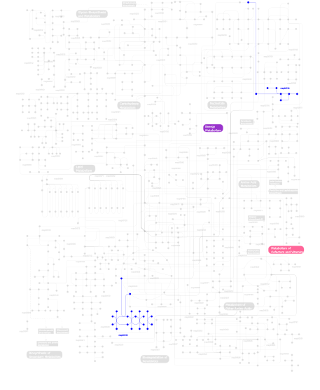
- Metabolism (metabolic pathways involving proteins which contain this domain)
- Structure (3D structures containing this domain)
- Links (links to other resources describing this domain)


