The domain within your query sequence starts at position 322 and ends at position 412; the E-value for the PKD domain shown below is 3.84e-1.
PKDRepeats in polycystic kidney disease 1 (PKD1) and other proteins |
|---|
| SMART accession number: | SM00089 |
|---|---|
| Description: | Polycystic kidney disease 1 protein contains 14 repeats, present elsewhere such as in microbial collagenases. |
| Interpro abstract (IPR022409): | The PKD (Polycystic Kidney Disease) domain was first identified in the Polycystic Kidney Disease protein, polycystin-1 (PDK1 gene), and contains an Ig-like fold consisting of a beta-sandwich of seven strands in two sheets with a Greek key topology, although some members have additional strands [ (PUBMED:9889186) ]. Polycystin-1 is a large cell-surface glycoprotein involved in adhesive protein-protein and protein-carbohydrate interactions; however it is not clear if the PKD domain mediates any of these interactions. PKD domains are also found in other proteins, usually in the extracellular parts of proteins involved in interactions with other proteins. For example, domains with a PKD-type fold are found in archaeal surface layer proteins that protect the cell from extreme environments [ (PUBMED:12377130) ], and in the human VPS10 domain-containing receptor SorCS2 [ (PUBMED:11499680) ]. |
| Family alignment: |
There are 95746 PKD domains in 42890 proteins in SMART's nrdb database.
Click on the following links for more information.
- Evolution (species in which this domain is found)
-
Taxonomic distribution of proteins containing PKD domain.
This tree includes only several representative species. The complete taxonomic breakdown of all proteins with PKD domain is also avaliable.
Click on the protein counts, or double click on taxonomic names to display all proteins containing PKD domain in the selected taxonomic class.
- Literature (relevant references for this domain)
-
Primary literature is listed below; Automatically-derived, secondary literature is also avaliable.
- Bateman A, Sandford R
- The PLAT domain: a new piece in the PKD1 puzzle.
- Curr Biol. 1999; 9: 58890-58890
- Sandford R et al.
- Comparative analysis of the polycystic kidney disease 1 (PKD1) gene reveals an integral membrane glycoprotein with multiple evolutionary conserved domains.
- Hum Mol Genet. 1997; 6: 1483-9
- Display abstract
PKD1 is the major locus of the common genetic disorder autosomal dominant polycystic kidney disease (ADPKD). Analysis of the predicted protein sequence of the human PKD1 gene, polycystin, shows a large molecule with a unique arrangement of extracellular domains and multiple putative transmembrane regions. The precise function of polycystin remains unclear with a paucity of mutations to define key structural and functional domains. To refine the structure of this protein we have cloned the genomic region encoding the Fugu PKD1 gene. Fugu PKD1 spans 36 kb of genomic DNA and has greater complexity with 54 exons compared with 46 in man. Comparative analysis of the predicted protein sequences shows a lower level of homology than in similar studies with identity of 40 and 59% similarity. However key structural motifs including leucine rich repeats (LRR), a C-type lectin and LDL-A like domains and 16 PKD repeats are maintained. A region of homology with the sea urchin REJ protein was also confirmed in Fugu but found to extend over 1000 amino acids. Several highly conserved intra- and extra-cellular regions, with no known sequence homologies, that are likely to be of functional importance were detected. The likely structure of the membrane associated region has been refined with similarity to the PKD2 protein and voltage gated Ca2+ and Na+ channels highlighted over part of this area. The overall protein structure has therefore been clarified and this comparative analysis derived structure will form the basis for the functional study of polycystin and its individual domains.
- Burn TC et al.
- Analysis of the genomic sequence for the autosomal dominant polycystic kidney disease (PKD1) gene predicts the presence of a leucine-rich repeat. The American PKD1 Consortium (APKD1 Consortium).
- Hum Mol Genet. 1995; 4: 575-82
- Display abstract
The complete genomic sequence of the gene responsible for the predominant form of polycystic kidney disease, PKD1, was determined to provide a framework for understanding the biology and evolution of the gene, and to aid in the development of molecular diagnostics. The DNA sequence of a 54 kb interval immediately upstream of the poly(A) addition signal sequence of the PKD1 transcript was determined, and then analyzed using computer methods. A leucine-rich repeat (LRR) motif was identified within the resulting predicted protein sequence of the PKD1 gene. By analogy with other LRR-containing proteins, this may explain some of the disease-related renal alterations such as mislocalization of membrane protein constituents and changes in the extracellular matrix organization. Finally, comparison of the genomic sequence and the published partial cDNA sequence showed several differences between the two sequences. The most significant difference detected predicts a novel carboxy-terminus for the PKD1 gene product.
- Hughes J et al.
- The polycystic kidney disease 1 (PKD1) gene encodes a novel protein with multiple cell recognition domains.
- Nat Genet. 1995; 10: 151-60
- Display abstract
Characterization of the polycystic kidney disease 1 (PKD1) gene has been complicated by genomic rearrangements on chromosome 16. We have used an exon linking strategy, taking RNA from a cell line containing PKD1 but not the duplicate loci, to clone a cDNA contig of the entire transcript. The transcript consists of 14,148 bp (including a correction to the previously described C terminus), distributed among 46 exons spanning 52 kb. The predicted PKD1 protein, polycystin, is a glycoprotein with multiple transmembrane domains and a cytoplasmic C-tail. The N-terminal extracellular region of over 2,500 aa contains leucine-rich repeats, a C-type lectin, 16 immunoglobulin-like repeats and four type III fibronectin-related domains. Our results indicate that polycystin is an integral membrane protein involved in cell-cell/matrix interactions.
- Metabolism (metabolic pathways involving proteins which contain this domain)
-

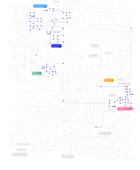
Click the image to view the interactive version of the map in iPath% proteins involved KEGG pathway ID Description 72.97  map00530
Aminosugars metabolism 5.41  map00500
Starch and sucrose metabolism 2.70 map00511 N-Glycan degradation 2.70  map00780
Biotin metabolism 2.70  map00860
Porphyrin and chlorophyll metabolism 2.70  map00310
Lysine degradation 2.70  map00561
Glycerolipid metabolism 2.70  map00051
Fructose and mannose metabolism 2.70 map02010 ABC transporters - General 2.70 map01032 Glycan structures - degradation This information is based on mapping of SMART genomic protein database to KEGG orthologous groups. Percentage points are related to the number of proteins with PKD domain which could be assigned to a KEGG orthologous group, and not all proteins containing PKD domain. Please note that proteins can be included in multiple pathways, ie. the numbers above will not always add up to 100%.
- Structure (3D structures containing this domain)
3D Structures of PKD domains in PDB
PDB code Main view Title 1b4r 
PKD DOMAIN 1 FROM HUMAN POLYCYSTEIN-1 1ctn 
CRYSTAL STRUCTURE OF A BACTERIAL CHITINASE AT 2.3 ANGSTROMS RESOLUTION 1edq 
CRYSTAL STRUCTURE OF CHITINASE A FROM S. MARCESCENS AT 1.55 ANGSTROMS 1ehn 
CRYSTAL STRUCTURE OF CHITINASE A MUTANT E315Q COMPLEXED WITH OCTA-N-ACETYLCHITOOCTAOSE (NAG)8. 1eib 
CRYSTAL STRUCTURE OF CHITINASE A MUTANT D313A COMPLEXED WITH OCTA-N-ACETYLCHITOOCTAOSE (NAG)8. 1ffq 
CRYSTAL STRUCTURE OF CHITINASE A COMPLEXED WITH ALLOSAMIDIN 1ffr 
CRYSTAL STRUCTURE OF CHITINASE A MUTANT Y390F COMPLEXED WITH HEXA-N-ACETYLCHITOHEXAOSE (NAG)6 1k9t 
Chitinase a complexed with tetra-N-acetylchitotriose 1l0q 
Tandem YVTN beta-propeller and PKD domains from an archaeal surface layer protein 1nh6 
Structure of S. marcescens chitinase A, E315L, complex with hexasaccharide 1rd6 
Crystal Structure of S. Marcescens Chitinase A Mutant W167A 1wgo 
Solution structure of the PKD domain from human VPS10 domain-containing receptor SorCS2 1x6l 
Crystal structure of S. marcescens chitinase A mutant W167A 1x6n 
Crystal structure of S. marcescens chitinase A mutant W167A in complex with allosamidin 2c26 
Structural basis for the promiscuous specificity of the carbohydrate- binding modules from the beta-sandwich super family 2c4x 
Structural basis for the promiscuous specificity of the carbohydrate- binding modules from the beta-sandwich super family 2kzw 
Solution NMR Structure of Q8PSA4 from Methanosarcina mazei, Northeast Structural Genomics Consortium Target MaR143A 2wk2 
Chitinase A from Serratia marcescens ATCC990 in complex with Chitotrio-thiazoline dithioamide. 2wly 
Chitinase A from Serratia marcescens ATCC990 in complex with Chitotrio-thiazoline. 2wlz 
Chitinase A from Serratia marcescens ATCC990 in complex with Chitobio- thiazoline. 2wm0 
Chitinase A from Serratia marcescens ATCC990 in complex with Chitobio- thiazoline thioamide. 2y3u 
Crystal structure of apo collagenase G from Clostridium histolyticum at 2.55 Angstrom resolution 2y50 
Crystal Structure of Collagenase G from Clostridium histolyticum at 2. 80 Angstrom Resolution 2y6i 
Crystal Structure of Collagenase G from Clostridium histolyticum in complex with Isoamylphosphonyl-Gly-Pro-Ala at 3.25 Angstrom Resolution 2y72 
Crystal structure of the PKD Domain of Collagenase G from Clostridium Histolyticum at 1.18 Angstrom Resolution. 2yrl 
Solution structure of the PKD domain from KIAA 1837 protein 3aro 
Crystal Structure Analysis of Chitinase A from Vibrio harveyi with novel inhibitors - apo structure 3arp 
Crystal Structure Analysis of Chitinase A from Vibrio harveyi with novel inhibitors - complex structure with DEQUALINIUM 3arq 
Crystal Structure Analysis of Chitinase A from Vibrio harveyi with novel inhibitors - complex structure with IDARUBICIN 3arr 
Crystal Structure Analysis of Chitinase A from Vibrio harveyi with novel inhibitors - complex structure with PENTOXIFYLLINE 3ars 
Crystal Structure Analysis of Chitinase A from Vibrio harveyi with novel inhibitors - apo structure of mutant W275G 3art 
Crystal Structure Analysis of Chitinase A from Vibrio harveyi with novel inhibitors - W275G mutant complex structure with DEQUALINIUM 3aru 
Crystal Structure Analysis of Chitinase A from Vibrio harveyi with novel inhibitors - W275G mutant complex structure with PENTOXIFYLLINE 3arv 
Crystal Structure Analysis of Chitinase A from Vibrio harveyi with novel inhibitors - complex structure with Sanguinarine 3arw 
Crystal Structure Analysis of Chitinase A from Vibrio harveyi with novel inhibitors - complex structure with chelerythrine 3arx 
Crystal Structure Analysis of Chitinase A from Vibrio harveyi with novel inhibitors - complex structure with Propentofylline 3ary 
Crystal Structure Analysis of Chitinase A from Vibrio harveyi with novel inhibitors - complex structure with 2-(imidazolin-2-yl)-5-isothiocyanatobenzofuran 3arz 
Crystal Structure Analysis of Chitinase A from Vibrio harveyi with novel inhibitors - complex structure with 2-(imidazolin-2-yl)-5-isothiocyanatobenzofuran 3as0 
Crystal Structure Analysis of Chitinase A from Vibrio harveyi with novel inhibitors - W275G mutant complex structure with Sanguinarine 3as1 
Crystal Structure Analysis of Chitinase A from Vibrio harveyi with novel inhibitors - W275G mutant complex structure with chelerythrine 3as2 
Crystal Structure Analysis of Chitinase A from Vibrio harveyi with novel inhibitors - W275G mutant complex structure with Propentofylline 3as3 
Crystal Structure Analysis of Chitinase A from Vibrio harveyi with novel inhibitors - W275G mutant complex structure with 2-(imidazolin-2-yl)-5-isothiocyanatobenzofuran 3b8s 
Crystal structure of wild-type chitinase A from Vibrio harveyi 3b9a 
Crystal structure of Vibrio harveyi chitinase A complexed with hexasaccharide 3b9d 
Crystal structure of Vibrio harveyi chitinase A complexed with pentasaccharide 3b9e 
Crystal structure of inactive mutant E315M chitinase A from Vibrio harveyi 4aqo 
CRYSTAL STRUCTURE OF THE CALCIUM BOUND PKD-like DOMAIN OF COLLAGENASE G FROM CLOSTRIDIUM HISTOLYTICUM AT 0.99 ANGSTROM RESOLUTION. 4jgu 
Crystal structure of Clostridium histolyticum colH collagenase collagen-binding domain 2b at 1.4 Angstrom resolution in presence of calcium 4jrw 
Crystal structure of Clostridium histolyticum colg collagenase PKD domain 2 at 1.6 Angstrom resolution 4l9d 
Crystal structure of the PKD1 domain from Vibrio cholerae metalloprotease PrtV 4tn9 
4TN9 4u6t 
4U6T 4u7k 
4U7K 5dez 
5DEZ 5df0 
5DF0 - Links (links to other resources describing this domain)
-
INTERPRO IPR022409 PFAM PKD

