The domain within your query sequence starts at position 2882 and ends at position 3037; the E-value for the SEC14 domain shown below is 2.08e-12.
VIEPYRRVISHGGYYGDGLNAIIVFAACFLPDSSRADYHYVMENLFLYVISTLELMVAED
YMIVYLNGATPRRKMPGLGWMKKCYQMIDRRLRKNLKSFIIVHPSWFIRTILAVTRPFIS
SKFSSKIKYVTSLSELSGLIPMDCIHIPESIIKYDE
SEC14Domain in homologues of a S. cerevisiae phosphatidylinositol transfer protein (Sec14p) |
|---|
| SMART accession number: | SM00516 |
|---|---|
| Description: | Domain in homologues of a S. cerevisiae phosphatidylinositol transfer protein (Sec14p) and in RhoGAPs, RhoGEFs and the RasGAP, neurofibromin (NF1). Lipid-binding domain. The SEC14 domain of Dbl is known to associate with G protein beta/gamma subunits. |
| Interpro abstract (IPR001251): | The CRAL-TRIO domain is a protein structural domain that binds small lipophilic molecules [ (PUBMED:12767229) ]. The domain is named after cellular retinaldehyde-binding protein (CRALBP) and TRIO guanine exchange factor. The CRAL-TRIO domain is found in GTPase-activating proteins (GAPs), guanine nucleotide exchange factors (GEFs) and a family of hydrophobic ligand binding proteins, including the yeast SEC14 protein and mammalian retinaldehyde- and alpha-tocopherol-binding proteins. The domain may either constitute all of the protein or only part of it [ (PUBMED:2198263) (PUBMED:8349655) (PUBMED:9461221) (PUBMED:10829015) ]. The structure of the domain in SEC14 proteins has been determined [ (PUBMED:9461221) ]. The structure contains several alpha helices as well as a beta sheet composed of 6 strands. Strands 2,3,4 and 5 form a parallel beta sheet with strands 1 and 6 being anti-parallel. The structure also identified a hydrophobic binding pocket for lipid binding. |
| Family alignment: |
There are 22196 SEC14 domains in 22009 proteins in SMART's nrdb database.
Click on the following links for more information.
- Evolution (species in which this domain is found)
-
Taxonomic distribution of proteins containing SEC14 domain.
This tree includes only several representative species. The complete taxonomic breakdown of all proteins with SEC14 domain is also avaliable.
Click on the protein counts, or double click on taxonomic names to display all proteins containing SEC14 domain in the selected taxonomic class.
- Literature (relevant references for this domain)
-
Primary literature is listed below; Automatically-derived, secondary literature is also avaliable.
- Aravind L, Neuwald AF, Ponting CP
- Sec14p-like domains in NF1 and Dbl-like proteins indicate lipid regulation of Ras and Rho signaling.
- Curr Biol. 1999; 9: 1957-1957
- Nishida K, Kaziro Y, Satoh T
- Association of the proto-oncogene product dbl with G protein betagamma subunits.
- FEBS Lett. 1999; 459: 186-90
- Display abstract
The Rho family of GTP-binding proteins has been implicated in the regulation of various cellular functions including actin cytoskeleton-dependent morphological change. Its activity is directed by intracellular signals mediated by various types of receptors such as G protein-coupled receptors. However, the mechanisms underlying receptor-dependent regulation of Rho family members remain incompletely understood. The guanine nucleotide exchange factor (GEF) Dbl targets Rho family proteins thereby stimulating their GDP/GTP exchange, and thus is believed to be involved in receptor-mediated regulation of the proteins. Here, we show the association of Dbl with G protein betagamma subunits (Gbetagamma) in transient co-expression and cell-free systems. An amino-terminal portion conserved among a subset of Dbl family proteins is sufficient for the binding of Gbetagamma. In fact, Ost and Kalirin, which contain this Gbetagamma-binding motif, also associate with Gbetagamma. c-Jun N-terminal kinase was synergistically activated upon co-expression of Dbl and Gbeta in a dominant-negative Rho-sensitive manner. However, GEF activity of Dbl toward Rho as measured by in vitro GDP binding assays remained unaffected following Gbetagamma binding, suggesting that additional signals may be required for the regulation of Dbl.
- Sha B, Luo M
- PI transfer protein: the specific recognition of phospholipids and its functions.
- Biochim Biophys Acta. 1999; 1441: 268-77
- Display abstract
Phosphatidylinositol transfer proteins (PITPs) can bind specifically and transfer a single phosphatidylinositol (PI) molecule between phospholipid membranes in an ATP-independent manner in vitro. PITPs exist in all the eukaryotic systems from yeast to human. PITP plays an essential role in intracellular vesicle flow and inositol lipid signaling. The crystal structure of yeast PITP Sec14p reveals a large hydrophobic pocket to accommodate the acyl chains of phospholipid molecules. At the opening of the pocket, a hydrogen bond network may render Sec14p the binding specificity to PI molecules. The structure suggests that the PI-binding ability may play an important role in the in vivo function of PITPs.
- Sha B, Phillips SE, Bankaitis VA, Luo M
- Crystal structure of the Saccharomyces cerevisiae phosphatidylinositol-transfer protein.
- Nature. 1998; 391: 506-10
- Display abstract
The yeast phosphatidylinositol-transfer protein (Sec14) catalyses exchange of phosphatidylinositol and phosphatidylcholine between membrane bilayers in vitro. In vivo, Sec14 activity is essential for vesicle budding from the Golgi complex. Here we report a three-dimensional structure for Sec14 at 2.5 A resolution. Sec14 consists of twelve alpha-helices, six beta-strands, eight 3(10)-helices and has two distinct domains. The carboxy-terminal domain forms a hydrophobic pocket which, in the crystal structure, is occupied by two molecules of n-octyl-beta-D-glucopyranoside and represents the phospholipid-binding domain. This pocket is reinforced by a string motif whose disruption in a sec14 temperature-sensitive mutant results in destabilization of the phospholipid-binding domain. Finally, we have identified an unusual surface helix that may play a critical role in driving Sec14-mediated phospholipid exchange. From this structure, we derive the first molecular clues into how a phosphatidylinositol-transfer protein functions.
- Steven R et al.
- UNC-73 activates the Rac GTPase and is required for cell and growth cone migrations in C. elegans.
- Cell. 1998; 92: 785-95
- Display abstract
unc-73 is required for cell migrations and axon guidance in C. elegans and encodes overlapping isoforms of 283 and 189 kDa that are closely related to the vertebrate Trio and Kalirin proteins, respectively. UNC-73A contains, in order, eight spectrin-like repeats, a Dbl/Pleckstrin homology (DH/PH) element, an SH3-like domain, a second DH/PH element, an immunoglobulin domain, and a fibronectin type III domain. UNC-73B terminates just downstream of the SH3-like domain. The first DH/PH element specifically activates the Rac GTPase in vitro and stimulates actin polymerization when expressed in Rat2 cells. Both functions are eliminated by introducing the S1216F mutation of unc-73(rh40) into this DH domain. Our results suggest that UNC-73 acts cell autonomously in a protein complex to regulate actin dynamics during cell and growth cone migrations.
- Alam MR, Johnson RC, Darlington DN, Hand TA, Mains RE, Eipper BA
- Kalirin, a cytosolic protein with spectrin-like and GDP/GTP exchange factor-like domains that interacts with peptidylglycine alpha-amidating monooxygenase, an integral membrane peptide-processing enzyme.
- J Biol Chem. 1997; 272: 12667-75
- Display abstract
Although the integral membrane proteins that catalyze steps in the biosynthesis of neuroendocrine peptides are known to contain routing information in their cytosolic domains, the proteins recognizing this routing information are not known. Using the yeast two-hybrid system, we previously identified P-CIP10 as a protein interacting with the cytosolic routing determinants of peptidylglycine alpha-amidating monooxygenase (PAM). P-CIP10 is a 217-kDa cytosolic protein with nine spectrin-like repeats and adjacent Dbl homology and pleckstrin homology domains typical of GDP/GTP exchange factors. In the adult rat, expression of P-CIP10 is most prevalent in the brain. Corticotrope tumor cells stably expressing P-CIP10 and PAM produce longer and more highly branched neuritic processes than nontransfected cells or cells expressing only PAM. The turnover of newly synthesized PAM is accelerated in cells co-expressing P-CIP10. P-CIP10 binds to selected members of the Rho subfamily of small GTP binding proteins (Rac1, but not RhoA or Cdc42). P-CIP10 (kalirin), a member of the Dbl family of proteins, may serve as part of a signal transduction system linking the catalytic domains of PAM in the lumen of the secretory pathway to cytosolic factors regulating the cytoskeleton and signal transduction pathways.
- Cerione RA, Zheng Y
- The Dbl family of oncogenes.
- Curr Opin Cell Biol. 1996; 8: 216-22
- Display abstract
Genetic screening and biochemical studies during the past few years have led to the discovery of a family of cell growth regulatory proteins and oncogene products for which the Dbl oncoprotein is a prototype. These putative guanine nucleotide exchange factors for Rho family small GTP-binding proteins (G proteins) all contain a Dbl homology domain in tandem with a pleckstrin homology domain, and seem to activate specific members of the Rho family of proteins to elicit various biological functions in cells. The Dbl homology domain is directly responsible for binding and activating the small G proteins to mediate downstream signaling events, whereas the pleckstrin homology domain may serve to target these positive regulators of G proteins to specific cellular locations to carry out the signaling task. Despite the increasing interest in the Dbl family of proteins, there is still a good deal to learn regarding the biochemical mechanisms that underlie their diverse biological functions.
- DelVecchio RL, Tonks NK
- Characterization of two structurally related Xenopus laevis protein tyrosine phosphatases with homology to lipid-binding proteins.
- J Biol Chem. 1994; 269: 19639-45
- Display abstract
We have chosen Xenopus laevis as a model system to study how protein tyrosine phosphatases (PTPases) function in growth and development. As an initial step, we have previously isolated in a polymerase chain reaction (PCR)-based protocol cDNA fragments which correspond to sequences within the catalytic domains of PTPases (Yang, Q., and Tonks, N. K. (1993) Adv. Protein Phosphatases 7, 359-372). Two of these PCR products, designated X1 and X10, have now been used to screen a X. laevis ovary cDNA library to obtain complete coding sequences for two distinct PTPases. The X1 cDNA encodes a protein (PTPX1) of 693 amino acids (approximately 79 kDa); the X10 cDNA encodes a protein of 597 amino acids (approximately 69 kDa). Both PTPX1 and PTPX10 lack potential membrane spanning sequences and therefore can be classified as non-transmembrane/cytoplasmic PTPases. While the overall structure of these PTPases are similar, sharing segments of 95% amino acid identity, they differ in that PTPX1 contains a unique 97-amino acid insert between the N-terminal segment and C-terminal catalytic domain. The absence of complete identity between PTPX1 and PTPX10 suggests that these two sequences are the products of separate genes and not the result of alternative splicing. This conclusion is confirmed by PCR analysis of Xenopus genomic DNA. Both PTPases share sequence identities in their N-terminal segments with two lipid-binding proteins, cellular retinaldehyde-binding protein and SEC14p, a phospholipid transferase. In addition, the unique insert sequence of PTPX1 shares identity with PSSA, a protein involved in phosphatidylserine biosynthesis. Sequence comparison suggests that PTPX10 is the Xenopus homolog of the human PTPase Meg-02, while PTPX1 is a structurally related yet distinct PTPase. Intrinsic PTPase activity of PTPX1 and PTPX10 was demonstrated in lysates of Sf9 cells infected with recombinant baculoviruses encoding either enzyme. PTPX1 can be recovered in both soluble and membrane fractions from Xenopus oocytes with the membrane form exhibiting approximately 4-fold higher activity than the soluble form.
- Gu M, Warshawsky I, Majerus PW
- Cloning and expression of a cytosolic megakaryocyte protein-tyrosine-phosphatase with sequence homology to retinaldehyde-binding protein and yeast SEC14p.
- Proc Natl Acad Sci U S A. 1992; 89: 2980-4
- Display abstract
Protein tyrosine phosphorylation is important in the regulation of cell growth, the cell cycle, and malignant transformation. We have cloned a cDNA that encodes a cytosolic protein-tyrosine-phosphatase (PTPase), MEG2, from MEG-01 cell and human umbilical vein endothelial cell cDNA libraries. The 4-kilobase cDNA sequence of PTPase MEG2 corresponds in length to the mRNA transcript detected by Northern blotting. The predicted open reading frame encodes a 68-kDa protein composed of 593 amino acids and has no apparent signal or transmembrane sequences, suggesting that it is a cytosolic protein. The C-terminal region has a PTPase catalytic domain that has 30-40% amino acid identity to other known PTPases. The N-terminal region has 254 amino acids that are 28% identical to cellular retinaldehyde-binding protein and 24% identical to yeast SEC14p, a protein that has phosphatidylinositol transfer activity and is required for protein secretion through the Golgi complex in yeast. Recombinant PTPase MEG2 expressed in Escherichia coli possesses PTPase activity. PTPase MEG2 mRNA was detected in 12 cell lines tested, which suggests that this phosphatase is widely expressed. The structure of PTPase MEG2 implies that a tyrosine phosphatase could participate in the transfer of hydrophobic ligands or in functions of the Golgi apparatus.
- Bankaitis VA, Aitken JR, Cleves AE, Dowhan W
- An essential role for a phospholipid transfer protein in yeast Golgi function.
- Nature. 1990; 347: 561-2
- Display abstract
Progression of proteins through the secretory pathway of eukaryotic cells involves a continuous rearrangement of macromolecular structures made up of proteins and phospholipids. The protein SEC14p is essential for transport of proteins from the yeast Golgi complex. Independent characterization of the SEC14 gene and the PIT1 gene, which encodes a phosphatidylinositol/phosphatidylcholine transfer protein in yeast, indicated that these two genes are identical. Phospholipid transfer proteins are a class of cytosolic proteins that are ubiquitous among eukaryotic cells and are distinguished by their ability to catalyse the exchange of phospholipids between membranes in vitro. We show here that the SEC14 and PIT1 genes are indeed identical and that the growth phenotype of a sec14-1ts mutant extends to the inability of its transfer protein to effect phospholipid transfer in vitro. These results therefore establish for the first time an in vivo function for a phospholipid transfer protein, namely a role in the compartment-specific stimulation of protein secretion.
- Disease (disease genes where sequence variants are found in this domain)
-
SwissProt sequences and OMIM curated human diseases associated with missense mutations within the SEC14 domain.
Protein Disease Neurofibromin (P21359) (SMART) OMIM:162200: Neurofibromatosis, type 1 ; Watson syndrome
OMIM:193520: Leukemia, juvenile myelomonocytic ; Melanoma, desmoplastic neurotropicAlpha-tocopherol transfer protein (P49638) (SMART) OMIM:600415: Ataxia with isolated vitamin E deficiency
OMIM:277460:Retinaldehyde-binding protein 1 (P12271) (SMART) OMIM:180090: Retinitis pigmentosa, autosomal recessive - Metabolism (metabolic pathways involving proteins which contain this domain)
-

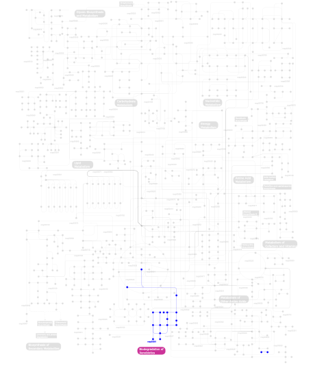
Click the image to view the interactive version of the map in iPath% proteins involved KEGG pathway ID Description 77.78 map04010 MAPK signaling pathway 11.11  map00361
gamma-Hexachlorocyclohexane degradation 11.11 map04610 Complement and coagulation cascades This information is based on mapping of SMART genomic protein database to KEGG orthologous groups. Percentage points are related to the number of proteins with SEC14 domain which could be assigned to a KEGG orthologous group, and not all proteins containing SEC14 domain. Please note that proteins can be included in multiple pathways, ie. the numbers above will not always add up to 100%.
- Structure (3D structures containing this domain)
3D Structures of SEC14 domains in PDB
PDB code Main view Title 1aua 
PHOSPHATIDYLINOSITOL TRANSFER PROTEIN SEC14P FROM SACCHAROMYCES CEREVISIAE 1o6u 
The Crystal Structure of Human Supernatant Protein Factor 1oip 
The Molecular Basis of Vitamin E Retention: Structure of Human Alpha-Tocopherol Transfer Protein 1oiz 
The Molecular Basis of Vitamin E Retention: Structure of Human Alpha-Tocopherol Transfer Protein 1olm 
Supernatant Protein Factor in Complex with RRR-alpha-Tocopherylquinone: A Link between Oxidized Vitamin E and Cholesterol Biosynthesis 1r5l 
Crystal Structure of Human Alpha-Tocopherol Transfer Protein Bound to its Ligand 2d4q 
Crystal structure of the Sec-PH domain of the human neurofibromatosis type 1 protein 2e2x 
Sec14 Homology Module of Neurofibromin in complex with phosphatitylethanolamine 3b74 
Crystal Structure of Yeast Sec14 Homolog Sfh1 in Complex with Phosphatidylethanolamine 3b7n 
Crystal Structure of Yeast Sec14 Homolog Sfh1 in Complex with Phosphatidylinositol 3b7q 
Crystal Structure of Yeast Sec14 Homolog Sfh1 in Complex with Phosphatidylcholine 3b7z 
Crystal Structure of Yeast Sec14 Homolog Sfh1 in Complex with Phosphatidylcholine or Phosphatidylinositol 3hx3 
Crystal structure of CRALBP mutant R234W 3hy5 
Crystal structure of CRALBP 3p7z 
Crystal structure of the Neurofibromin Sec14-PH module containing the patient derived mutation I1584V 3peg 
Crystal structure of Neurofibromins Sec14-PH module containing a patient derived duplication (TD) 3pg7 
Crystal structure of the H. sapiens NF1 SEC-PH domain (del1750 mutant) 3q8g 
Resurrection of a functional phosphatidylinositol transfer protein from a pseudo-Sec14 scaffold by directed evolution 3w67 
Crystal structure of mouse alpha-tocopherol transfer protein in complex with alpha-tocopherol and phosphatidylinositol-(3,4)-bisphosphate 3w68 
Crystal structure of mouse alpha-tocopherol transfer protein in complex with alpha-tocopherol and phosphatidylinositol-(4,5)-bisphosphate 4ciz 
Crystal structure of the complex of the Cellular Retinal Binding Protein with 9-cis-retinal 4cj6 
Crystal structure of the complex of the Cellular Retinal Binding Protein Mutant R234W with 9-cis-retinal 4fmm 
Dimeric Sec14 family homolog 3 from Saccharomyces cerevisiae presents some novel features of structure that lead to a surprising "dimer-monomer" state change induced by substrate binding 4j7p 
Crystal structure of Saccharomyces cerevisiae Sfh3 4j7q 
Crystal structure of Saccharomyces cerevisiae Sfh3 complexed with phosphatidylinositol 4m8z 
4M8Z 4omj 
4OMJ 4omk 
4OMK 4tlg 
4TLG 4uyb 
4UYB - Links (links to other resources describing this domain)
-
INTERPRO IPR001251

